==============================================
⬇️✨⬇️🎉⬇️🔥⬇️📚⬇️
Click the link to purchase on Amazon 🎉📚
==============================================
🎥 Check Out All Videos at Once! 📺
👉 Visit Visualizing MSK Blog to explore a wide range of videos! 🩻
https://visualizingmsk.blogspot.com/?view=magazine
📚 You can also find them on MSK MRI Blog and Naver Blog! 📖
https://www.instagram.com/msk_mri/
Click now to stay updated with the latest content! 🔍✨
==============================================
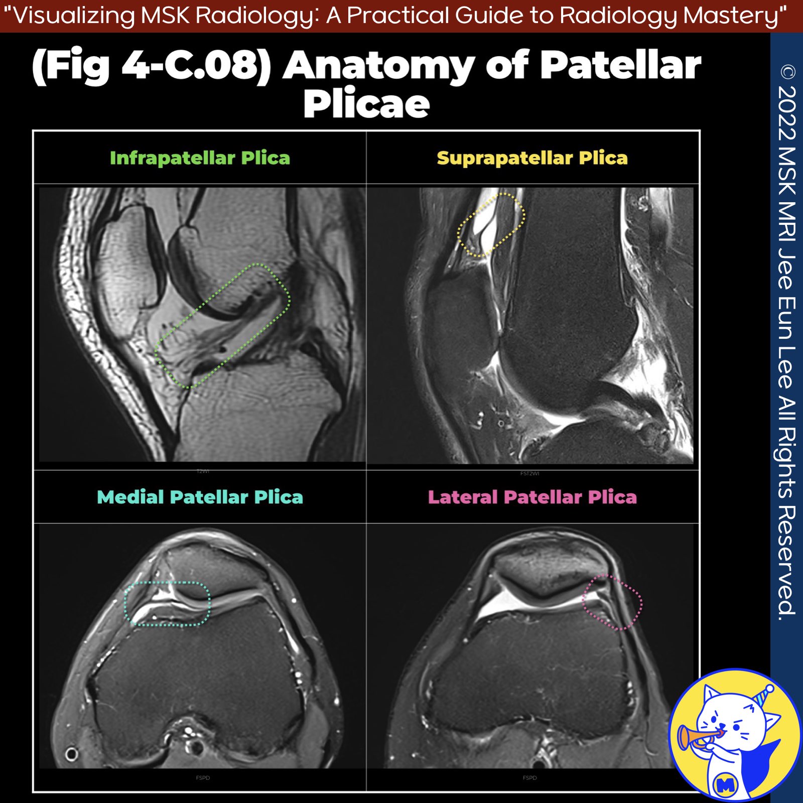
📌 Anatomy of the Plicae
- Plicae are thin folds of vascularized synovial tissue resulting from incomplete resorption of embryonic septa that partition the knee joint into medial, lateral, and suprapatellar compartments during development.
- There are four potential plicae, observed in descending order of frequency: infrapatellar plica, suprapatellar plica, mediopatellar plica, and lateral patellar plica . Most plicae are asymptomatic and incidentally seen at imaging or arthroscopy.
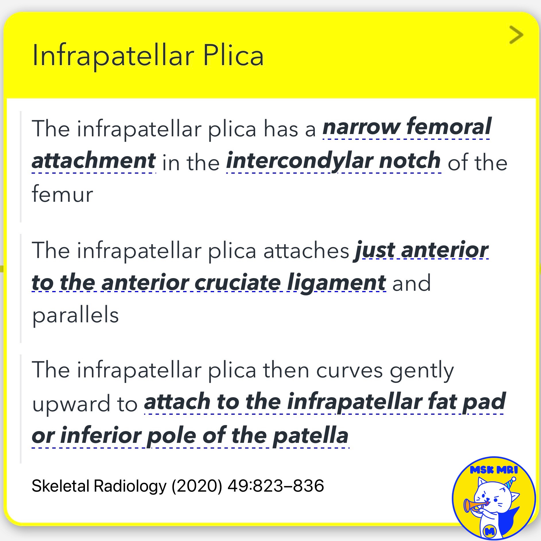
1️⃣ Infrapatellar Plica
- The infrapatellar plica has a narrow femoral attachment in the intercondylar notch of the femur, which attaches just anterior to and parallels the anterior cruciate ligament.
- It then curves gently upward to attach to the infrapatellar fat pad or the inferior pole of the patella.
- This plica is a remnant of the septum between the medial and lateral tibiofemoral compartments .
2️⃣ Suprapatellar Plica
- Also known as the superior plica, plica synovialis suprapatellaris, superomedial plica, or medial suprapatellar plica, the suprapatellar plica runs from the anterior aspect of the femoral metaphysis to the posterior aspect of the quadriceps tendon, inserting above the patella .
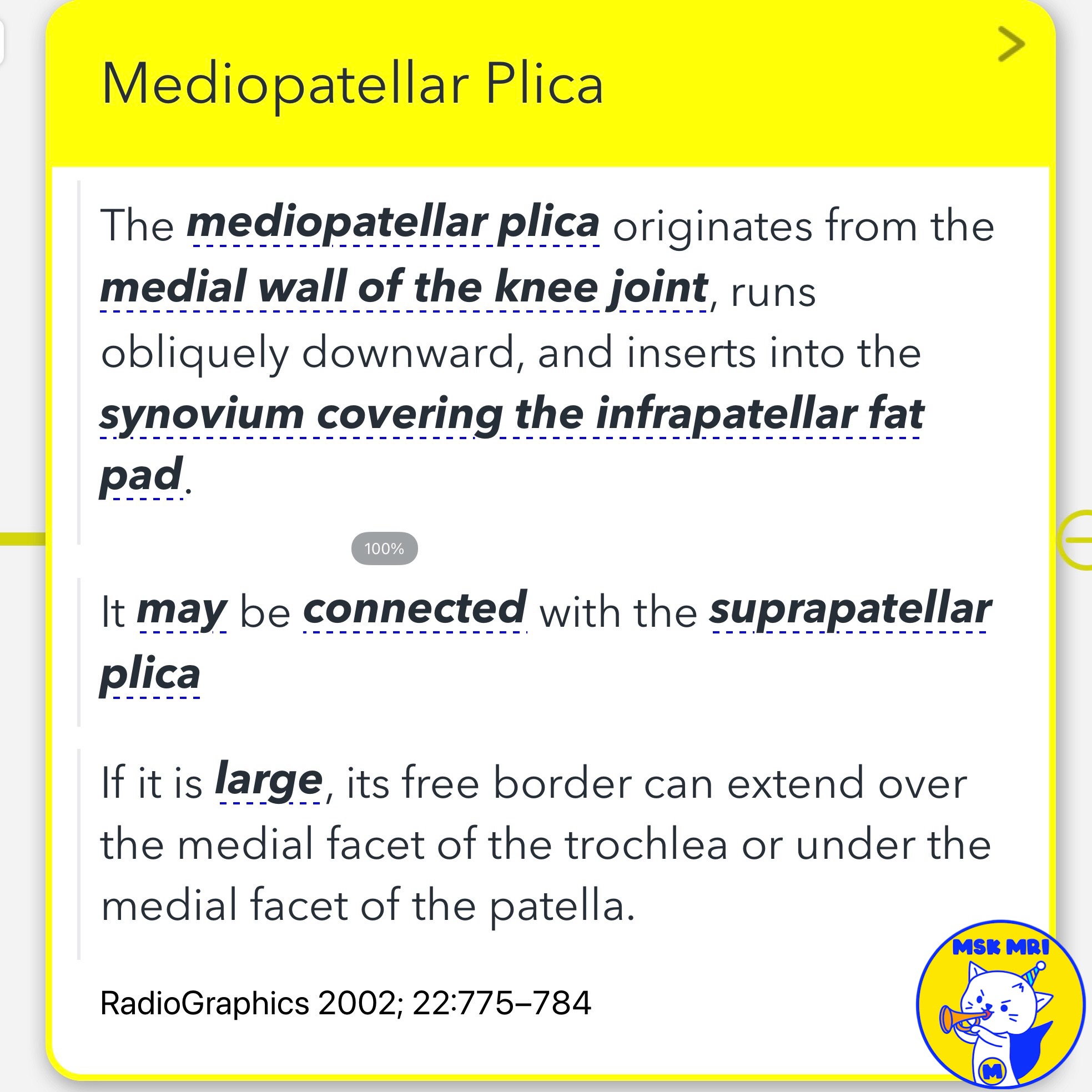
3️⃣ Mediopatellar Plica
- The mediopatellar plica originates from the medial wall of the knee joint, runs obliquely downward, and inserts into the synovium covering the infrapatellar fat pad.
- It may be connected with the suprapatellar plica.
- If it is large, its free border can extend over the medial facet of the trochlea or under the medial facet of the patella .
4️⃣ Lateral Patellar Plica
- The lateral patellar plica is the least common of the four knee plicae.
- Lateral plicae originate from the lateral joint capsule, coursing in the lateral gutter of the knee, and attaching to the infrapatellar fat pad.
- It should be distinguished from other synovial folds in the lateral gutter, such as the lateral alar fold, superolateral fold, and transverse synovial arcuate fold .
References
- RadioGraphics 2018; 38:2069–2101
- Skeletal Radiol. 2018 Aug;47(8):1069-1086
- Skeletal Radiology (2020) 49:823–836
- Skeletal Radiol. 2018 Aug;47(8):1069-1086
- RadioGraphics 2002; 22:775–784
- MRI Web Clinic — November 2018 Synovial Plicae of the Knee
"Visualizing MSK Radiology: A Practical Guide to Radiology Mastery"
© 2022 MSK MRI Jee Eun Lee All Rights Reserved.
No unauthorized reproduction, redistribution, or use for AI training.
#Plicae #KneeAnatomy #SynovialTissue #InfrapatellarPlica #SuprapatellarPlica #MediopatellarPlica #LateralPatellarPlica #Radiology #MedicalImaging #Orthopedics


'✅ Knee MRI Mastery > Chap 4BCD. Anterior knee' 카테고리의 다른 글
| (Fig 4-C.10) Sakakibara Classification (0) | 2024.06.19 |
|---|---|
| (Fig 4-C.09) Medial Plica Syndrome (0) | 2024.06.19 |
| (Fig 4-C.07) Peri-cruciate Fat Pad Inflammation (0) | 2024.06.18 |
| (Fig 4-C.06) Quadriceps Fat Pad Edema (0) | 2024.06.18 |
| (Fig 4-C.05) Pre-femoral Fat Pad (0) | 2024.06.17 |