==============================================
⬇️✨⬇️🎉⬇️🔥⬇️📚⬇️
Click the link to purchase on Amazon 🎉📚
==============================================
🎥 Check Out All Videos at Once! 📺
👉 Visit Visualizing MSK Blog to explore a wide range of videos! 🩻
https://visualizingmsk.blogspot.com/?view=magazine
📚 You can also find them on MSK MRI Blog and Naver Blog! 📖
https://www.instagram.com/msk_mri/
Click now to stay updated with the latest content! 🔍✨
==============================================
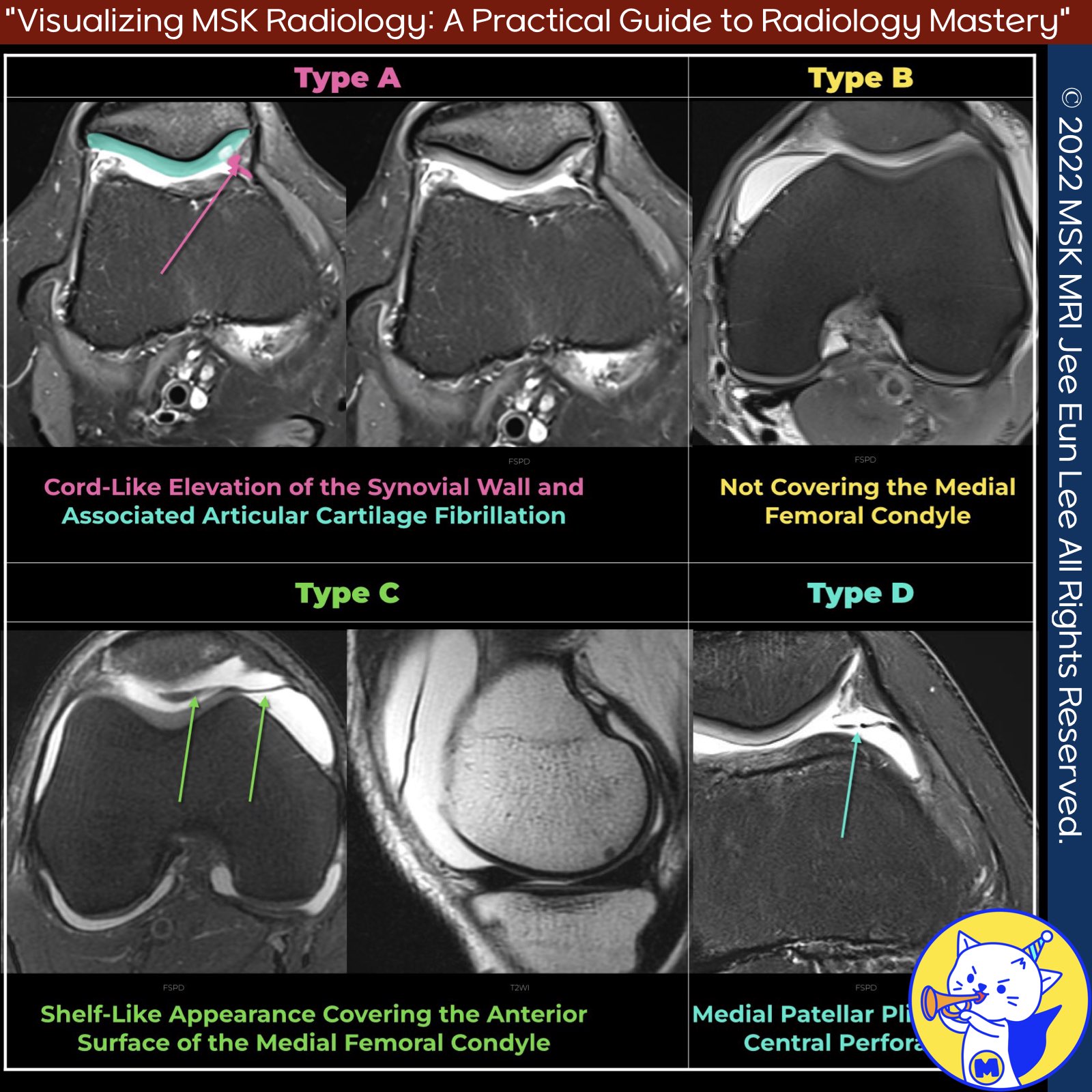
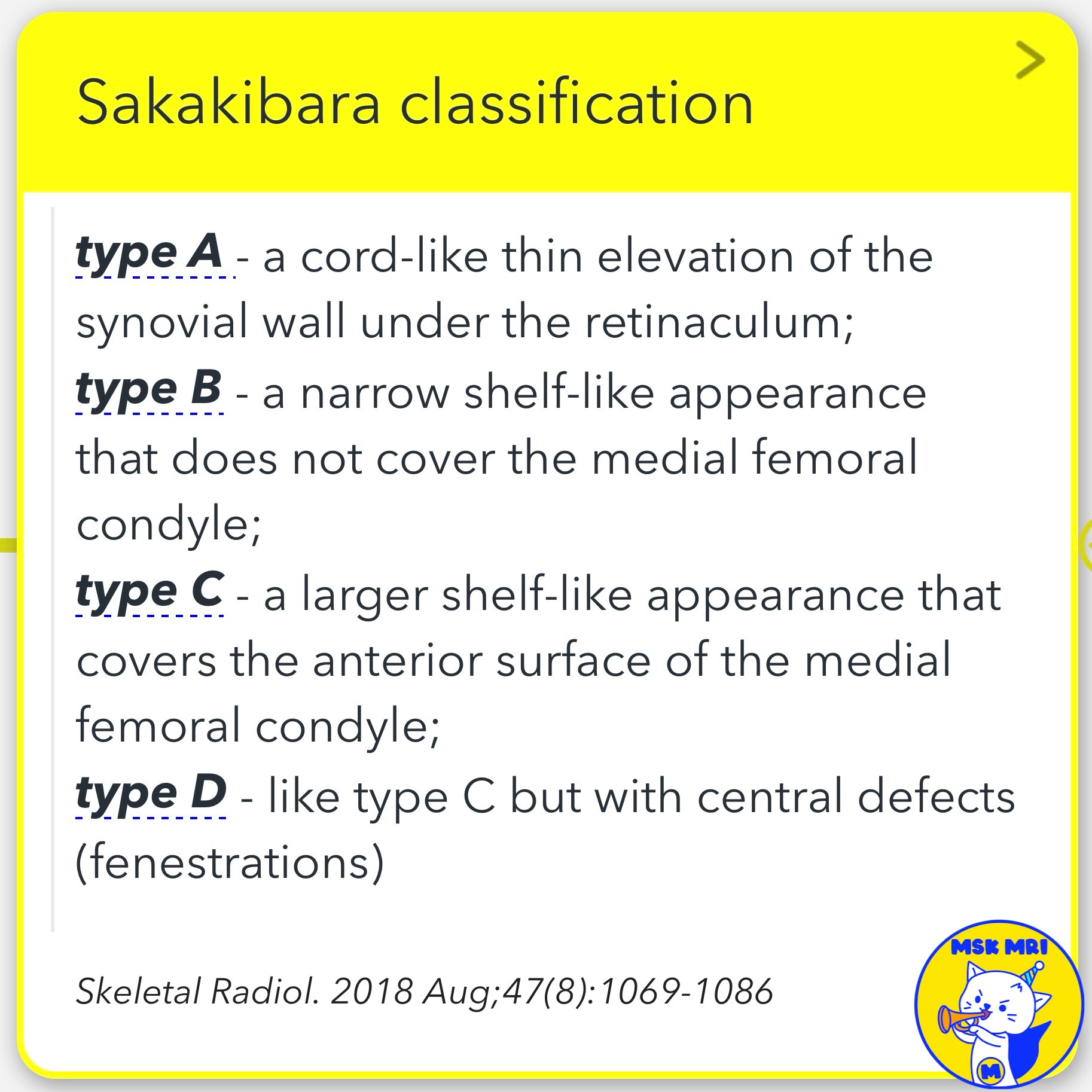
📌 Sakakibara Classification
- The Sakakibara classification is a system used to describe medial patellar plicae based on their size and morphology.
- This classification aids in identifying the potential for symptomatic issues related to the plicae.
✅ Type A
- Type A is characterized by a cord-like thin elevation of the synovial wall under the retinaculum.
- This type does not cover the medial patellofemoral articular surface and is less likely to cause symptoms.
✅ Type B
- Type B presents a narrow shelf-like appearance that does not cover the medial femoral condyle.
- Similar to Type A, Type B plicae are smaller and less likely to be symptomatic.
✅ Type C
- Type C is a larger shelf-like plica that covers the anterior surface of the medial femoral condyle.
- This type extends to cover the articular surfaces, making it more likely to cause symptoms.
- MRI findings often show a wide plica extending beyond the medial margin of the trochlear articular surface, corresponding to a Sakakibara Type C lesion.
✅ Type D
- Type D is similar to Type C but includes central defects known as fenestrations.
- This type also covers the articular surfaces and is more frequently associated with symptoms, but it is relatively rare.
- MRI findings for Type D lesions include thickening and increased intrasubstance signal on T2-weighted images, fenestration, focal fluid adjacent to the plica, and interposition between the patella and femoral trochlea on multiple contiguous images.
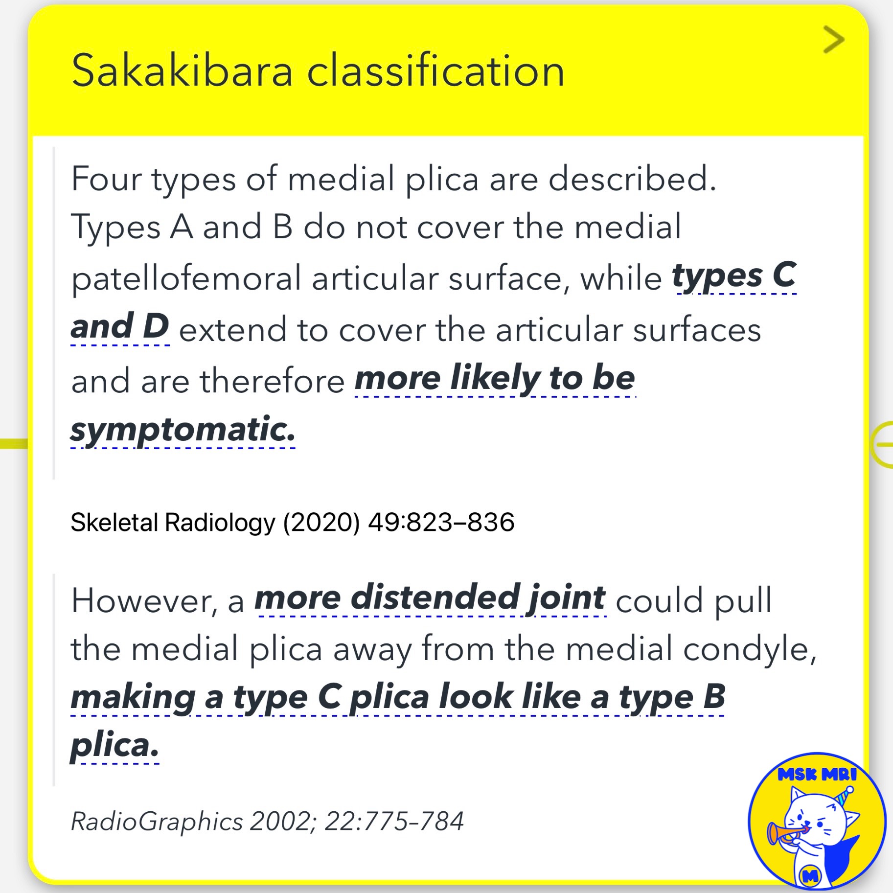
★ Clinical Significance
- Types A and B, which do not cover the medial patellofemoral articular surface, are generally smaller and less likely to cause symptoms.
- In contrast, Types C and D, which extend to cover the articular surfaces, are more prone to being symptomatic due to their size and position.
- Additionally, a more distended joint could pull the medial plica away from the medial condyle, making a Type C plica appear like a Type B plica.
References
- Skeletal Radiol. 2018 Aug;47(8):1069-1086.
- Skeletal Radiology (2020) 49:823–836.
- RadioGraphics 2002; 22:775–784.
- MRI Web Clinic — November 2018 Synovial Plicae of the Knee.
"Visualizing MSK Radiology: A Practical Guide to Radiology Mastery"
© 2022 MSK MRI Jee Eun Lee All Rights Reserved.
No unauthorized reproduction, redistribution, or use for AI training.
#SakakibaraClassification, #MedialPlica, #KneeMRI, #Radiology, #KneeInjury, #Orthopedics, #MedicalImaging, #SynovialPlica, #Musculoskeletal, #KneePain


'✅ Knee MRI Mastery > Chap 4BCD. Anterior knee' 카테고리의 다른 글
| (Fig 4-C.12) Acute Injury of Infrapatellar Plica (0) | 2024.06.21 |
|---|---|
| (Fig 4-C.11) Suprapatellar Plica Causing Compartmentalization (0) | 2024.06.19 |
| (Fig 4-C.09) Medial Plica Syndrome (0) | 2024.06.19 |
| (Fig 4-C.08) Anatomy of Patellar Plicae (0) | 2024.06.18 |
| (Fig 4-C.07) Peri-cruciate Fat Pad Inflammation (0) | 2024.06.18 |