==============================================
⬇️✨⬇️🎉⬇️🔥⬇️📚⬇️
Click the link to purchase on Amazon 🎉📚
==============================================
🎥 Check Out All Videos at Once! 📺
👉 Visit Visualizing MSK Blog to explore a wide range of videos! 🩻
https://visualizingmsk.blogspot.com/?view=magazine
📚 You can also find them on MSK MRI Blog and Naver Blog! 📖
https://www.instagram.com/msk_mri/
Click now to stay updated with the latest content! 🔍✨
==============================================
📌 Suprapatellar Plica
- The suprapatellar plica, a membrane remnant from embryonic development, is found in around 16% of adults.
✅ Symptoms
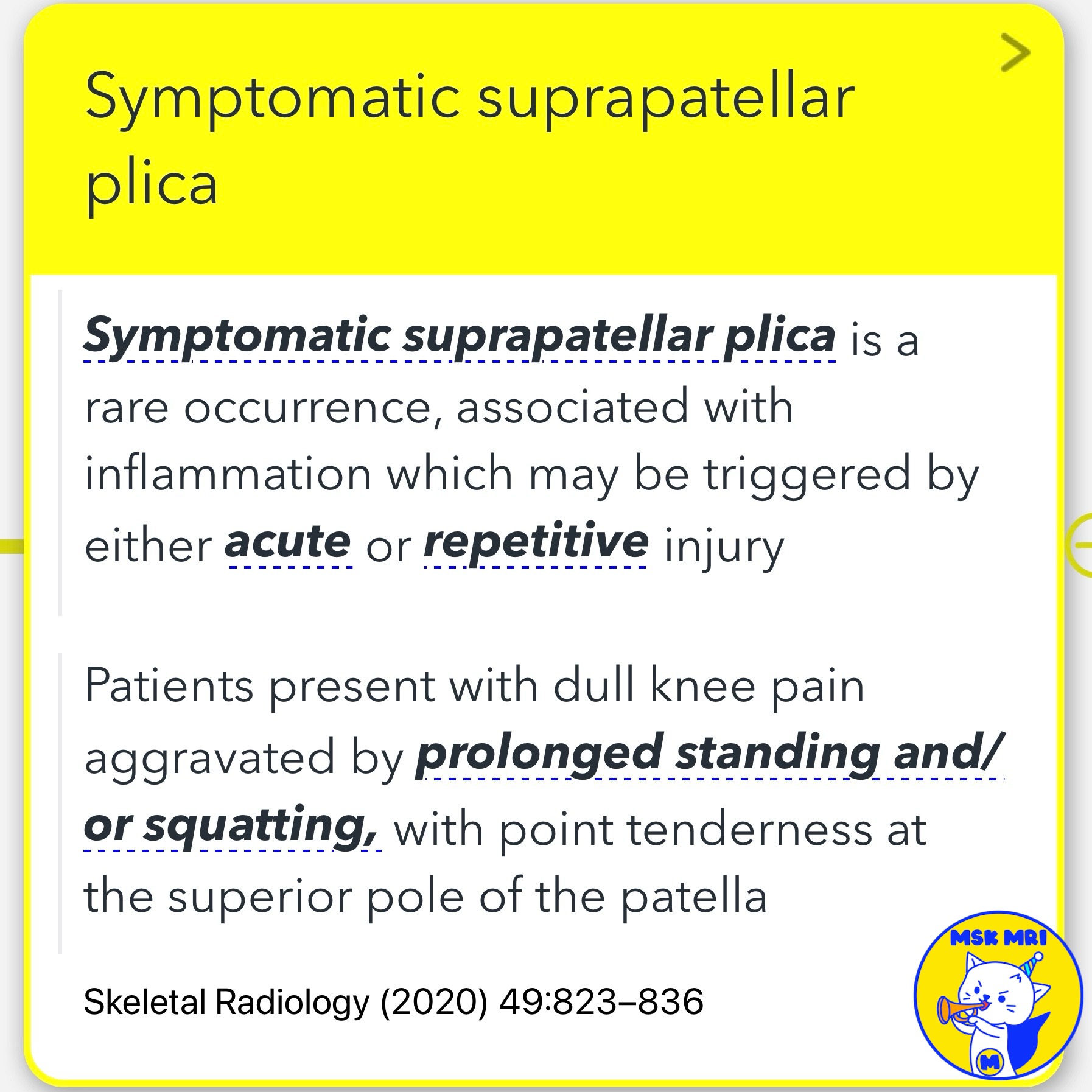
- Symptomatic suprapatellar plica is rare and results from inflammation due to injury.
- Patients report dull knee pain worsened by standing or squatting and tenderness at the top of the patella.
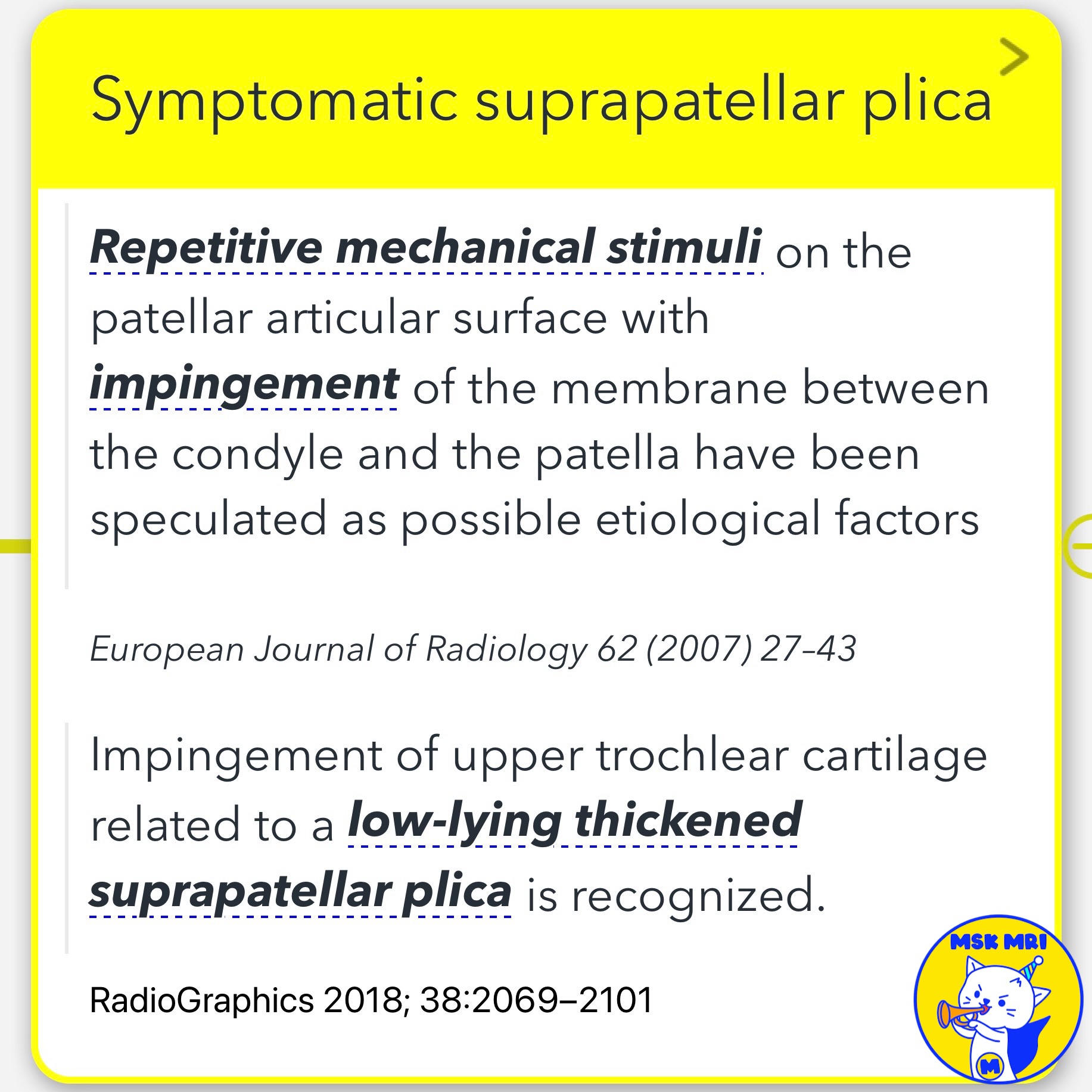
- Possible causes include repetitive mechanical stress on the patellar surface and impingement between the condyle and patella.
✅ Impingement Issues
- A low-lying, thickened plica can impinge on the upper trochlear cartilage, leading to secondary chondromalacia from compression during knee flexion.
✅ Bursa Compartmentalization
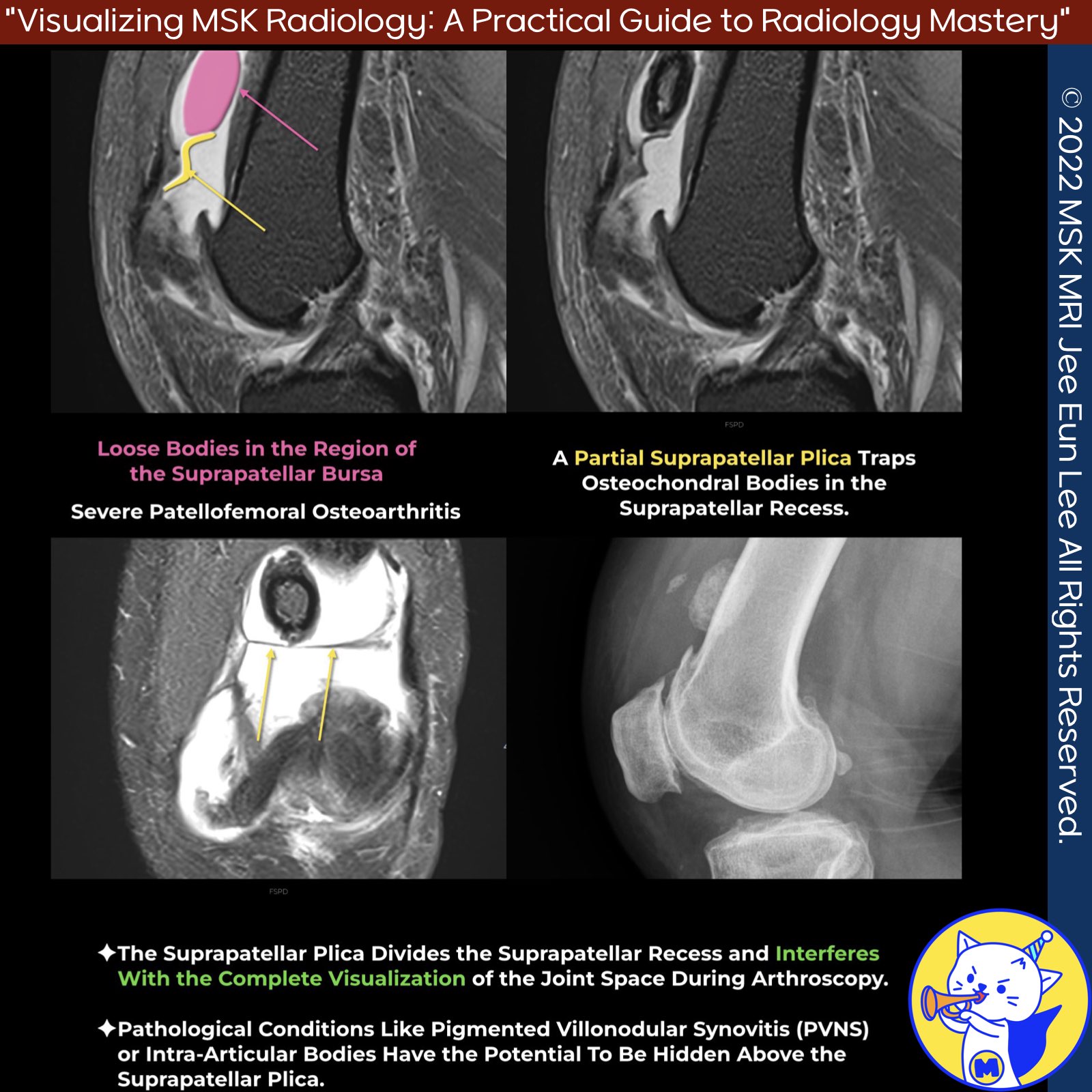
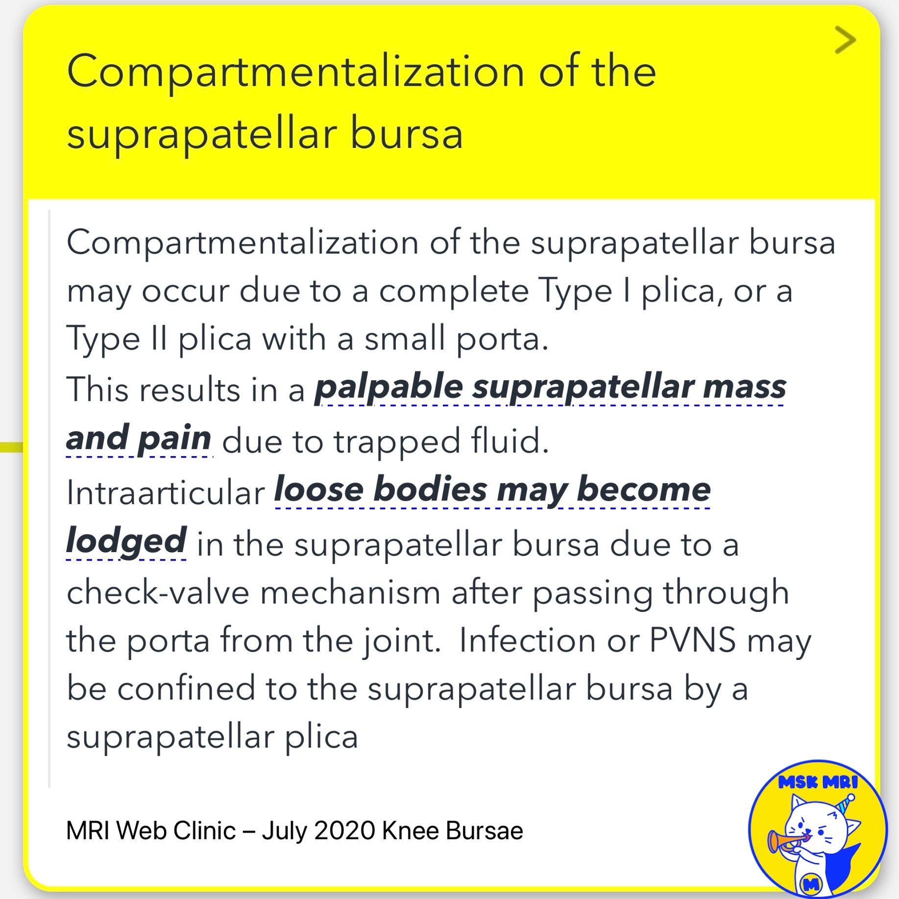
- Complete or partially open plica can compartmentalize the suprapatellar bursa, causing a palpable mass and pain from a trapped fluid.
- This may also trap loose bodies, exacerbating symptoms.
- A complete septum plica can obscure joint space visualization during arthroscopy, potentially hiding conditions like PVNS or intra-articular bodies.
References
- Skeletal Radiology (2020) 49:823–836
- European Journal of Radiology 62 (2007) 27–43
- RadioGraphics 2018; 38:2069–2101
- MRI Web Clinic – July 2020 Knee Bursae
- Skeletal Radiol. 2018 Aug;47(8):1069-1086
"Visualizing MSK Radiology: A Practical Guide to Radiology Mastery"
© 2022 MSK MRI Jee Eun Lee All Rights Reserved.
No unauthorized reproduction, redistribution, or use for AI training.
#SuprapatellarPlica, #KneePain, #OrthopedicRadiology, #KneeInjury, #JointCompartmentalization, #KneeBursae, #PlicaSyndrome, #Chondromalacia, #Arthroscopy, #RadiologyResearch


'✅ Knee MRI Mastery > Chap 4BCD. Anterior knee' 카테고리의 다른 글
| (Fig 4-C.13) Posterior Hoffa's Fat Pad Impingement, Plica syndrome (0) | 2024.06.21 |
|---|---|
| (Fig 4-C.12) Acute Injury of Infrapatellar Plica (0) | 2024.06.21 |
| (Fig 4-C.10) Sakakibara Classification (0) | 2024.06.19 |
| (Fig 4-C.09) Medial Plica Syndrome (0) | 2024.06.19 |
| (Fig 4-C.08) Anatomy of Patellar Plicae (0) | 2024.06.18 |