==============================================
⬇️✨⬇️🎉⬇️🔥⬇️📚⬇️
Click the link to purchase on Amazon 🎉📚
==============================================
🎥 Check Out All Videos at Once! 📺
👉 Visit Visualizing MSK Blog to explore a wide range of videos! 🩻
https://visualizingmsk.blogspot.com/?view=magazine
📚 You can also find them on MSK MRI Blog and Naver Blog! 📖
https://www.instagram.com/msk_mri/
Click now to stay updated with the latest content! 🔍✨
==============================================
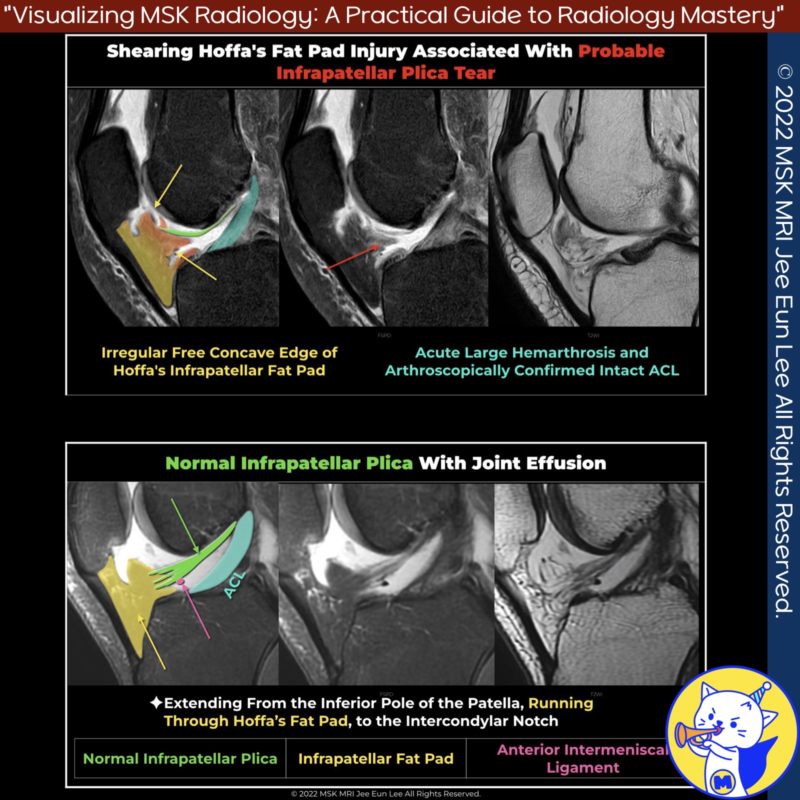
📌 Acute Injury of Infrapatellar Plica
- Acute injury to the infrapatellar plica can cause hemarthrosis, pain, and a locking sensation in the knee.
- These symptoms usually follow trauma or repetitive knee motion.
✅ MRI Findings
- Sprain: Increased signal along the infrapatellar plica (IPP) on fluid-sensitive sequences, less than fluid signal intensity[1].
- Tear: Increased signal along the IPP with fluid signal intensity on fluid-sensitive sequences[1].
- Edema: Edema within the infrapatellar fat pad in a characteristic curvilinear pattern along the course of the plica[2].
- Hoffa’s Fat Pad: Tears appear as a linear high signal intensity zone on fat-suppressed T2-weighted images, not corresponding to normal Hoffa’s clefts[3]. Scars appear as low signal-intensity tissue on all sequences[3].
✅Differential Diagnosis
- Synovial Clefts: Two synovium-lined clefts in Hoffa’s fat pad, one vertical and one horizontal, are normally present. These clefts are smoothly marginated and follow a continuous curvilinear pattern[1].
- Sprain vs. Tear: Sprain or tear hyperintensity typically has ragged or irregular margins, unlike the smooth margins of synovial clefts[1].
- Horizontal Synovial Clefts: Differentiated from an IPP tear by their continuous nature with the joint cavity and roof formation by the IPP[1].
✅ Associated Injuries
- Infrapatellar plica tears often coexist with other knee injuries, especially ACL tears.
- Edema and scarring of Hoffa’s fat pad are common in knees with torn ACLs due to instability and fat pad impingement around the ligamentum mucosum.
- Abnormalities in Hoffa’s fat pad, such as focal and diffuse edema, tears, scars, and synovial proliferation, are more frequent in knees with ACL injuries[4].
References
- SA J Radiol. 2021 Feb 19;25(1):1973
- MRI Web Clinic — November 2018 Synovial Plicae of the Knee
- Skeletal Radiol (2008) 37:301–306
- Skeletal Radiology (2020) 49:823–836
"Visualizing MSK Radiology: A Practical Guide to Radiology Mastery"
© 2022 MSK MRI Jee Eun Lee All Rights Reserved.
No unauthorized reproduction, redistribution, or use for AI training.
#InfrapatellarPlica #KneeInjury #MRI #SynovialPlicae #HoffasFatPad #ACLTear #KneePain #Hemarthrosis #FatPadEdema #Radiology


'✅ Knee MRI Mastery > Chap 4BCD. Anterior knee' 카테고리의 다른 글
| (Fig 4-C.14) Medial Synovial Fold of the PCL (0) | 2024.06.21 |
|---|---|
| (Fig 4-C.13) Posterior Hoffa's Fat Pad Impingement, Plica syndrome (0) | 2024.06.21 |
| (Fig 4-C.11) Suprapatellar Plica Causing Compartmentalization (0) | 2024.06.19 |
| (Fig 4-C.10) Sakakibara Classification (0) | 2024.06.19 |
| (Fig 4-C.09) Medial Plica Syndrome (0) | 2024.06.19 |