Click the link to purchase on Amazon 🎉📚
==============================================
🎥 Check Out All Videos at Once! 📺
👉 Visit Visualizing MSK Blog to explore a wide range of videos! 🩻
https://visualizingmsk.blogspot.com/?view=magazine
📚 You can also find them on MSK MRI Blog and Naver Blog! 📖
https://www.instagram.com/msk_mri/
Click now to stay updated with the latest content! 🔍✨
==============================================
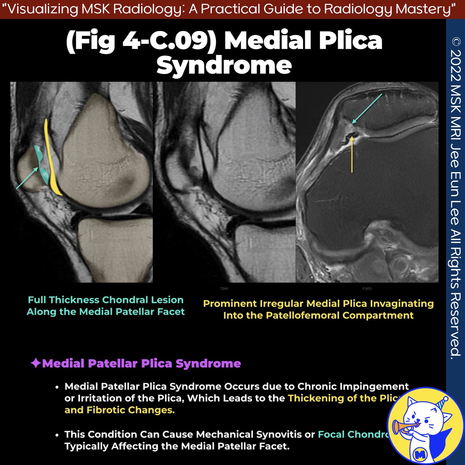
📌 Medial Patellar Plica Syndrome
- Medial patellar plica syndrome arises when the medial plica of the knee becomes thickened and fibrotic due to chronic impingement or irritation, leading to mechanical synovitis or focal chondrosis, particularly involving the medial patellar facet (1).
✅ Pathophysiology
- The medial plica is the most commonly symptomatic plica in the knee. Inflammation or fibrosis causes loss of elasticity, interrupting its gliding action and leading to irritation at the anterior medial knee (2).
- Thickened and fibrotic plicae often have irregular margins, making them less flexible and more likely to cause mechanical symptoms during joint movement, such as snapping over the medial femoral trochlea and patella (3).
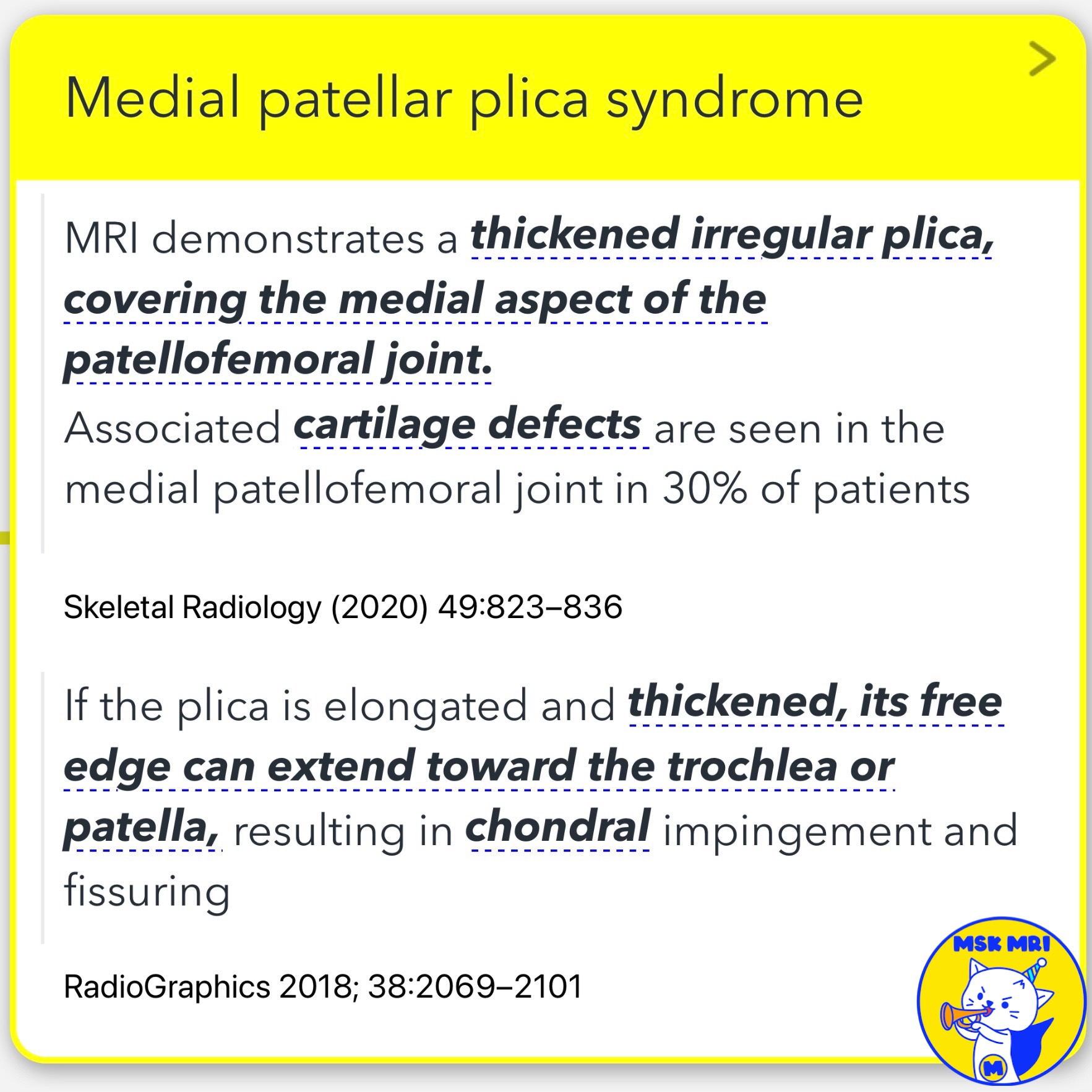
- If the plica is elongated and thickened, its free edge can extend toward the trochlea or patella, causing chondral impingement and fissuring (3).
✅ Clinical Features
Symptoms of medial patellar plica syndrome include:
- Pain worsened by activity
- Swelling
- Sensations of locking or instability
- Rarely, a palpable cord-like structure in the medial peripatellar area (3).
As the disease progresses, inflammation can lead to articular cartilage loss and traction on the adjacent synovium, exacerbating patient symptoms (4).
✅ Diagnosis
- Diagnosis is primarily based on clinical findings. However, MRI can reveal a thickened, irregular plica covering the medial aspect of the patellofemoral joint.
- In approximately 30% of patients, associated cartilage defects are visible in the medial patellofemoral joint (5).
References
- Skeletal Radiol. 2018 Aug;47(8):1069-1086.
- MRI Web Clinic — November 2018 Synovial Plicae of the Knee.
- RadioGraphics 2018; 38:2069–2101.
- MRI Web Clinic — November 2018 Synovial Plicae of the Knee.
- Skeletal Radiology (2020) 49:823–836.
"Visualizing MSK Radiology: A Practical Guide to Radiology Mastery"
© 2022 MSK MRI Jee Eun Lee All Rights Reserved.
No unauthorized reproduction, redistribution, or use for AI training.
#MedialPatellarPlicaSyndrome, #KneePain, #PlicaSyndrome, #Orthopedics, #KneeInjury, #SynovialPlicae, #SportsMedicine, #MRI, #ChondralImpingement, #JointHealth
'✅ Knee MRI Mastery > Chap 4BCD. Anterior knee' 카테고리의 다른 글
| (Fig 4-C.11) Suprapatellar Plica Causing Compartmentalization (0) | 2024.06.19 |
|---|---|
| (Fig 4-C.10) Sakakibara Classification (0) | 2024.06.19 |
| (Fig 4-C.08) Anatomy of Patellar Plicae (0) | 2024.06.18 |
| (Fig 4-C.07) Peri-cruciate Fat Pad Inflammation (0) | 2024.06.18 |
| (Fig 4-C.06) Quadriceps Fat Pad Edema (0) | 2024.06.18 |


