Click the link to purchase on Amazon 🎉📚
==============================================
🎥 Check Out All Videos at Once! 📺
👉 Visit Visualizing MSK Blog to explore a wide range of videos! 🩻
https://visualizingmsk.blogspot.com/?view=magazine
📚 You can also find them on MSK MRI Blog and Naver Blog! 📖
https://www.instagram.com/msk_mri/
Click now to stay updated with the latest content! 🔍✨
==============================================
📌 Ruptured Baker’s Cyst
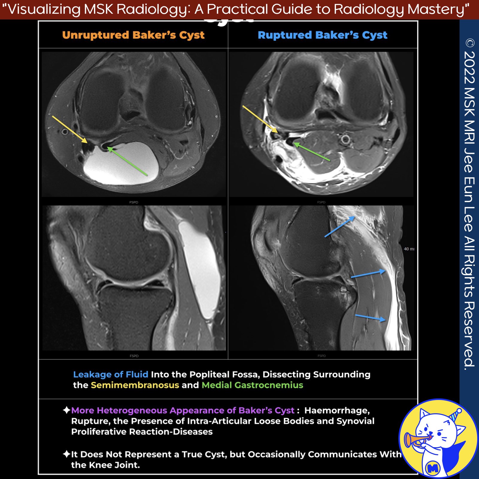
A ruptured Baker’s cyst, also known as a popliteal cyst, is efficiently demonstrated on MRI as a high signal intensity edema dispersing into the adjacent soft tissues and fascial planes on fat-suppressed T2-weighted sequences.
✅ Complications
1️⃣ Dissection: The cyst typically dissects inferomedially but can also dissect proximally, anteriorly, intermuscularly, or intramuscularly.
- Presentation: A dissecting Baker’s cyst may present with tender fullness at the posteromedial aspect of the knee just below the joint. These lesions represent rare instances where a Baker’s cyst enters through the muscle fascia and is present both outside and within a muscle compartment.
- Normally, a Baker’s cyst enlarges in the direction of least resistance, most commonly along the medial gastrocnemius muscle belly distally. If a focal fascial defect occurs, or at a pre-existing weak region, the Baker’s cyst can enter the muscle compartment.
2️⃣ Rupture: Leaking of cyst fluid into the popliteal fossa, between fascial planes, and surrounding the hamstrings and medial gastrocnemius muscles. This is accompanied by edema of the soft tissue and irregularity of the cyst wall .
3️⃣ Compression: The enlarged cyst can compress the popliteal vessels and tibial nerve, leading to vascular and neurological symptoms.
4️⃣ Compartment Syndrome: The cyst can cause compartment syndrome, which can be either anterior or posterior.
✅ Clinical Presentation
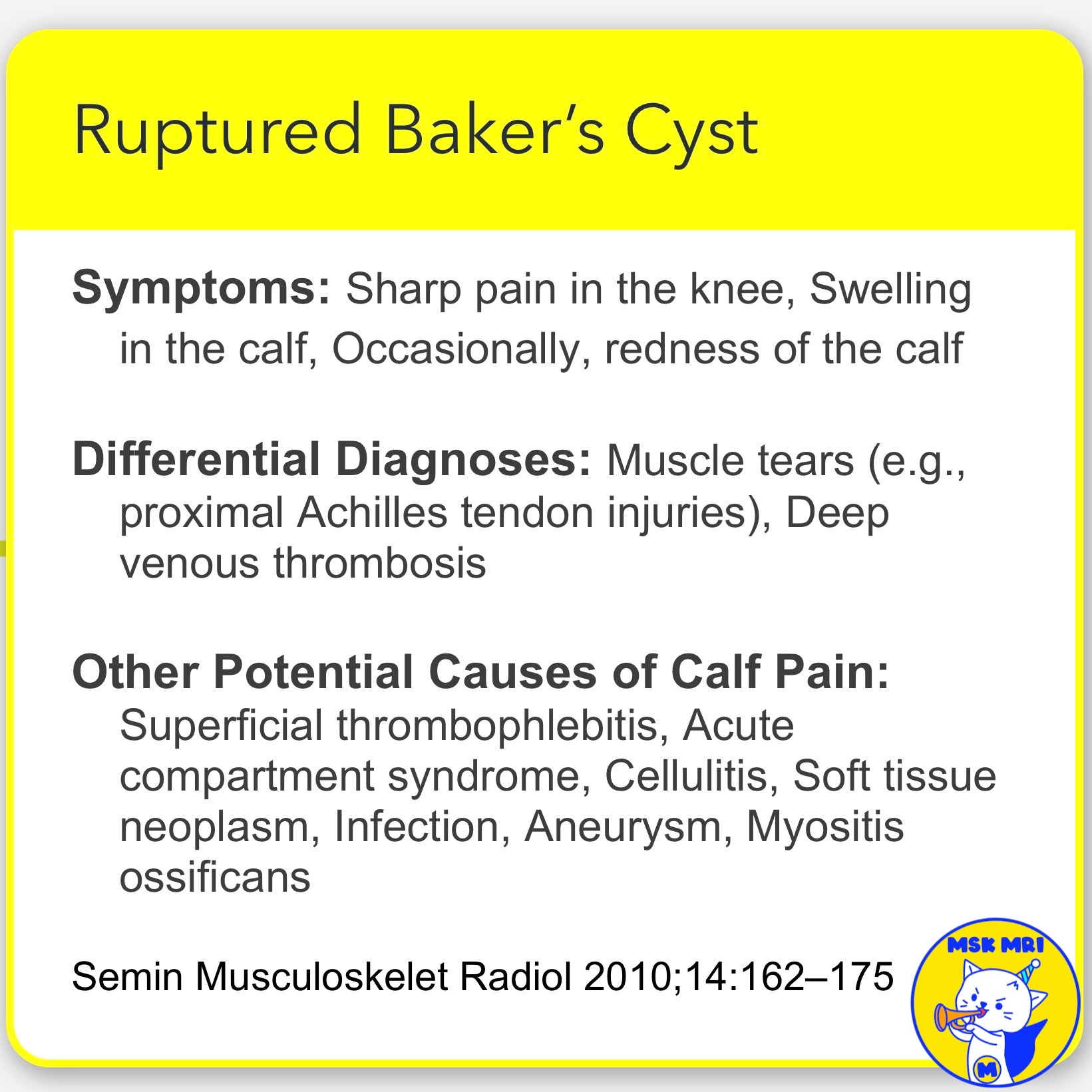
- Symptoms: Sharp pain in the knee, Swelling in the calf, Occasionally, redness of the calf
- Differential Diagnoses: Muscle tears (e.g., proximal Achilles tendon injuries), Deep venous thrombosis
- Other Potential Causes of Calf Pain: Superficial thrombophlebitis, Acute compartment syndrome, Cellulitis, Soft tissue neoplasm, Infection, Aneurysm, Myositis ossificans
References
- Insights Imaging (2013) 4:257–272
- MRI Web Clinic — September 2013 Proximal Gastrocnemius Tendon Pathology
- https://radiopaedia.org/articles/baker-cyst-2?lang=us
- Semin Musculoskelet Radiol 2010;14:162–175
"Visualizing MSK Radiology: A Practical Guide to Radiology Mastery"
© 2022 MSK MRI Jee Eun Lee All Rights Reserved.
No unauthorized reproduction, redistribution, or use for AI training.
#RupturedBakersCyst, #PoplitealCyst, #MRI, #SoftTissueEdema, #Gastrocnemius, #KneePain, #Dissection, #CompartmentSyndrome, #Compression, #Radiology
'✅ Knee MRI Mastery > Chap 4BCD. Anterior knee' 카테고리의 다른 글
| (Fig 4-D.11) Intramuscular Extension of Baker's Cyst (0) | 2024.06.25 |
|---|---|
| (Fig 4-D.10) Proximal and Distal Popliteal Cyst Dissection and Rupture (0) | 2024.06.24 |
| (Fig 4-D.08) Unruptured Baker’s Cyst (1) | 2024.06.24 |
| (Fig 4-D.07) Comparison of Parameniscal Cysts and Infrapatellar Ganglion Cysts (0) | 2024.06.23 |
| (Fig 4-D.06) Hoffa’s Fat Pad Ganglion Cysts (0) | 2024.06.23 |


