==============================================
⬇️✨⬇️🎉⬇️🔥⬇️📚⬇️
Click the link to purchase on Amazon 🎉📚
==============================================
🎥 Check Out All Videos at Once! 📺
👉 Visit Visualizing MSK Blog to explore a wide range of videos! 🩻
https://visualizingmsk.blogspot.com/?view=magazine
📚 You can also find them on MSK MRI Blog and Naver Blog! 📖
https://www.instagram.com/msk_mri/
Click now to stay updated with the latest content! 🔍✨
==============================================
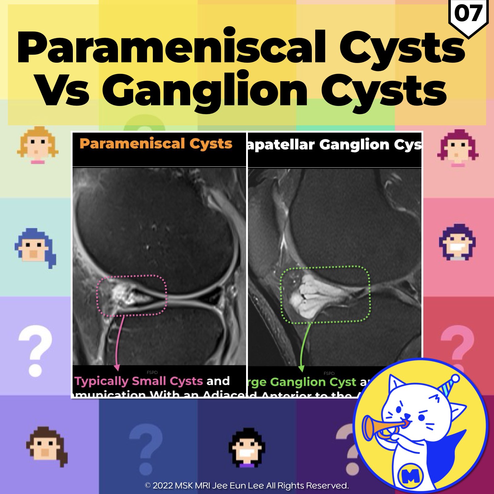
📌 Hoffa’s Fat Pad Ganglion Cysts and Parameniscal Cysts
✅ Hoffa’s Fat Pad Ganglion Cysts
- Hoffa’s fat pad ganglion cysts are most commonly found in the Hoffa fat pad, typically adjacent to the anterior horn of the lateral meniscus .
- On MR imaging, ganglia appear as well-defined, uni- or multi-loculated, fluid-like T2 hyperintense lesions.
- Depending on their protein content, ganglia may be hypo- or isointense on T1-weighted sequences .
- An uncomplicated ganglion appears as a well-defined homogeneous non-enhancing fluid-filled collection on MRI.
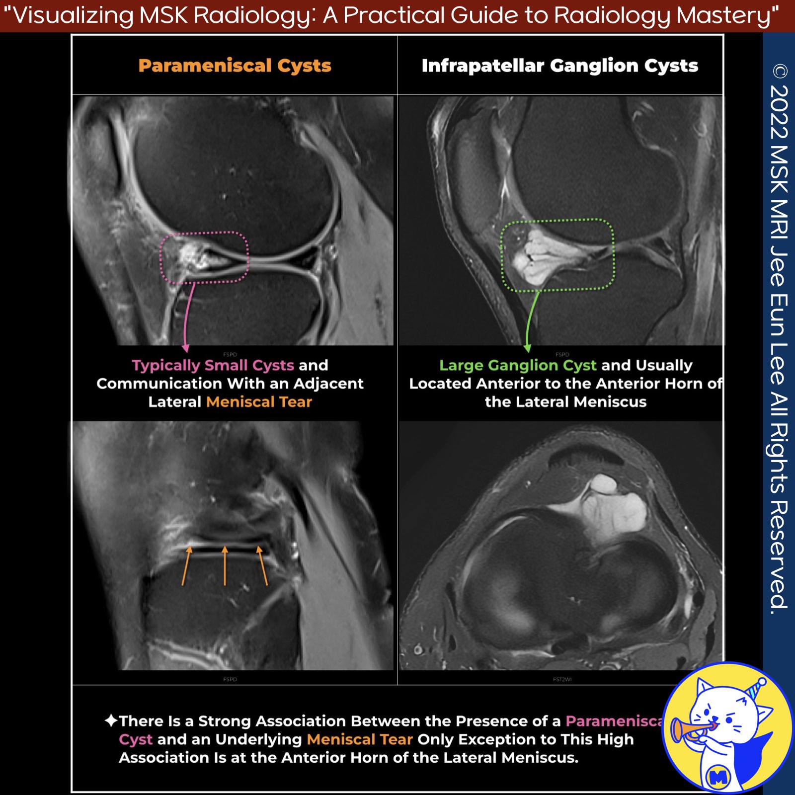
✅ Parameniscal Cysts
- On T2-weighted imaging, a parameniscal cyst is a high-signal-intensity fluid collection either directly overlying a meniscus or adjacent to a meniscus with a fluid track connecting to the periphery of a meniscus.
- There is a strong association between the presence of a parameniscal cyst and an underlying meniscal tear.
- The reported association between parameniscal cysts and meniscal tears has ranged from 90% to 100% in MRI series, except at the anterior horn of the lateral meniscus, where an underlying meniscal tear was found in only 64% of patients with these parameniscal cysts.
✅ Differences Between Ganglion Cysts and Parameniscal Cysts
- A parameniscal cyst can simulate a ganglion but is typically smaller and associated with an underlying meniscal tear.
- Confirming a parameniscal cyst is based on depicting a communication with an adjacent meniscal tear on any pulse sequence.
- Synovial fluid extrusion secondary to complex and horizontal meniscal tears (lateral more than medial) results in meniscal cyst formation, which may project into Hoffa’s fat pad.
References
- RadioGraphics 2018; 38:2069–2101
- Osteoarthritis Cartilage. 2016 Mar;24(3):383-97
- Indian J Radiol Imaging 2021;31:961–974
- AJR 2012; 199:481–499
"Visualizing MSK Radiology: A Practical Guide to Radiology Mastery"
© 2022 MSK MRI Jee Eun Lee All Rights Reserved.
No unauthorized reproduction, redistribution, or use for AI training.
#Radiology, #MRI, #GanglionCysts, #ParameniscalCysts, #HoffasFatPad, #MeniscalTears, #T2Imaging, #SynovialFluid, #JointSpace, #MedicalImaging


'✅ Knee MRI Mastery > Chap 4BCD. Anterior knee' 카테고리의 다른 글
| (Fig 4-D.09) Ruptured Baker’s Cyst (0) | 2024.06.24 |
|---|---|
| (Fig 4-D.08) Unruptured Baker’s Cyst (1) | 2024.06.24 |
| (Fig 4-D.06) Hoffa’s Fat Pad Ganglion Cysts (0) | 2024.06.23 |
| (Fig 4-D.05) Deep Infrapatellar Bursitis (0) | 2024.06.23 |
| (Fig 4-D.04) Morel-Lavallée Lesion (0) | 2024.06.23 |