Click the link to purchase on Amazon 🎉📚
==============================================
🎥 Check Out All Videos at Once! 📺
👉 Visit Visualizing MSK Blog to explore a wide range of videos! 🩻
https://visualizingmsk.blogspot.com/?view=magazine
📚 You can also find them on MSK MRI Blog and Naver Blog! 📖
https://www.instagram.com/msk_mri/
Click now to stay updated with the latest content! 🔍✨
==============================================
📌 Hoffa’s Fat Pad Ganglion Cysts
- Ganglion cysts at the anterior knee are well-marginated masses filled with gelatinous material lacking synovial lining.
- They are most commonly found in the Hoffa fat pad, typically adjacent to the anterior horn of the lateral meniscus .
- The mass can be large and expand the fat pad, resulting in symptoms of intra-articular impingement or limited motion.
✅ MRI Characteristics
- On MR imaging, ganglia appear as well-defined, uni- or multi-loculated, fluid-like T2 hyperintense lesions.
- Depending on their protein content, ganglia may be hypo- or isointense on T1-weighted sequences.
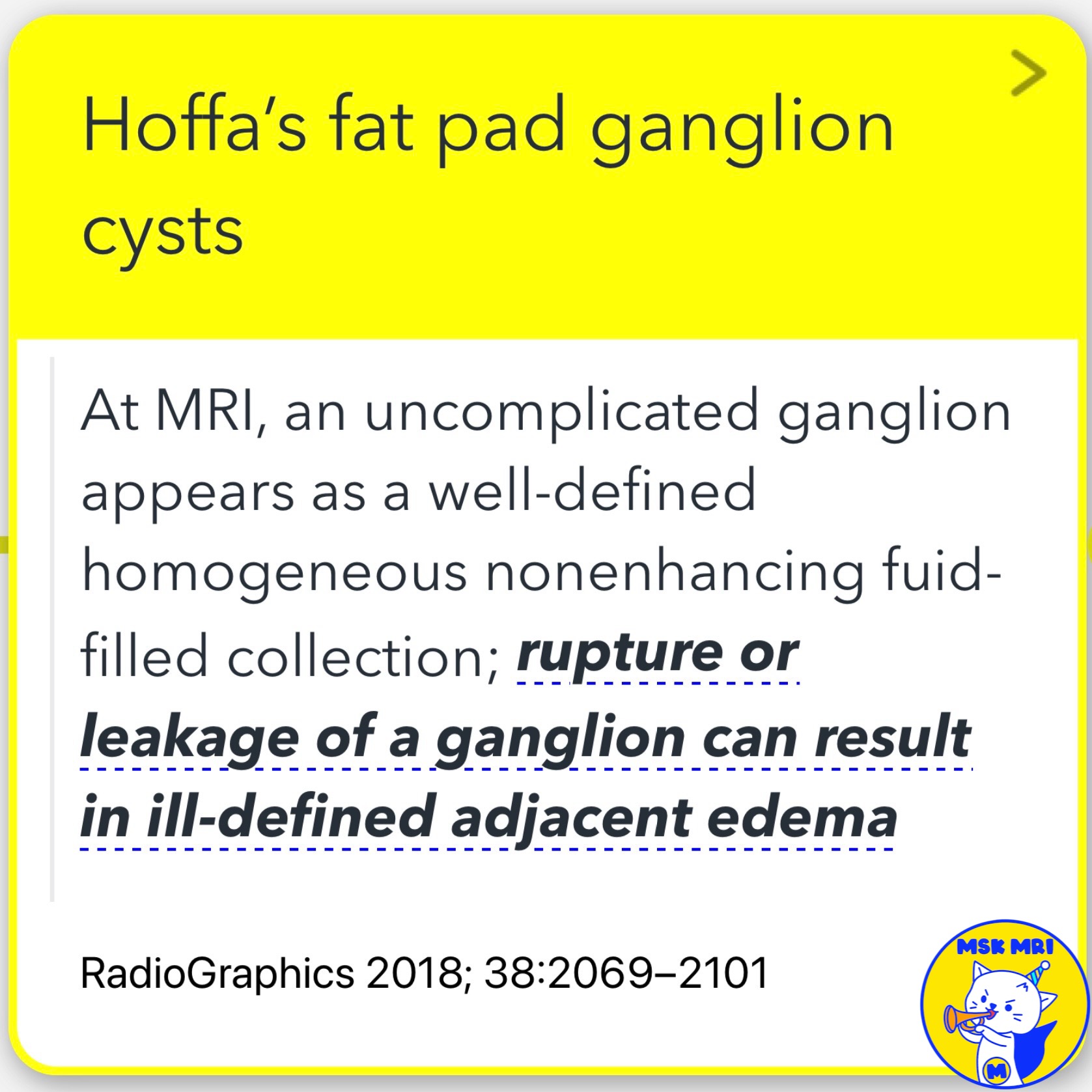
- An uncomplicated ganglion appears as a well-defined, homogeneous, non-enhancing fluid-filled collection on MRI.
- Rupture or leakage of a ganglion can result in ill-defined adjacent edema.
- Ganglion cysts can be solitary or multiple, round or globular, multi- or uniloculated, and may demonstrate a neck or communication with adjacent joint spaces .
✅Differential Diagnosis
- An intrahoffatic lesion or a synovial lesion such as haemangioma or synovial sarcoma may be misinterpreted as a ganglion cyst within the fat pad.
References
- RadioGraphics 2018; 38:2069–2101
- Osteoarthritis Cartilage. 2016 Mar;24(3):383-97
- Indian J Radiol Imaging 2021;31:961–974
- Insights Imaging (2013) 4:257–272
"Visualizing MSK Radiology: A Practical Guide to Radiology Mastery"
© 2022 MSK MRI Jee Eun Lee All Rights Reserved.
No unauthorized reproduction, redistribution, or use for AI training.
#HoffasFatPad #GanglionCysts #KneeMRI #Radiology #MusculoskeletalRadiology #KneePain #IntraArticularImpingement #MRImaging #KneeInjuries #GanglionCharacteristics
'✅ Knee MRI Mastery > Chap 4BCD. Anterior knee' 카테고리의 다른 글
| (Fig 4-D.08) Unruptured Baker’s Cyst (1) | 2024.06.24 |
|---|---|
| (Fig 4-D.07) Comparison of Parameniscal Cysts and Infrapatellar Ganglion Cysts (0) | 2024.06.23 |
| (Fig 4-D.05) Deep Infrapatellar Bursitis (0) | 2024.06.23 |
| (Fig 4-D.04) Morel-Lavallée Lesion (0) | 2024.06.23 |
| (Fig 4-D.03) Septic Prepatellar Bursitis (0) | 2024.06.23 |


