==============================================
⬇️✨⬇️🎉⬇️🔥⬇️📚⬇️
Click the link to purchase on Amazon 🎉📚
==============================================
🎥 Check Out All Videos at Once! 📺
👉 Visit Visualizing MSK Blog to explore a wide range of videos! 🩻
https://visualizingmsk.blogspot.com/?view=magazine
📚 You can also find them on MSK MRI Blog and Naver Blog! 📖
https://www.instagram.com/msk_mri/
Click now to stay updated with the latest content! 🔍✨
==============================================
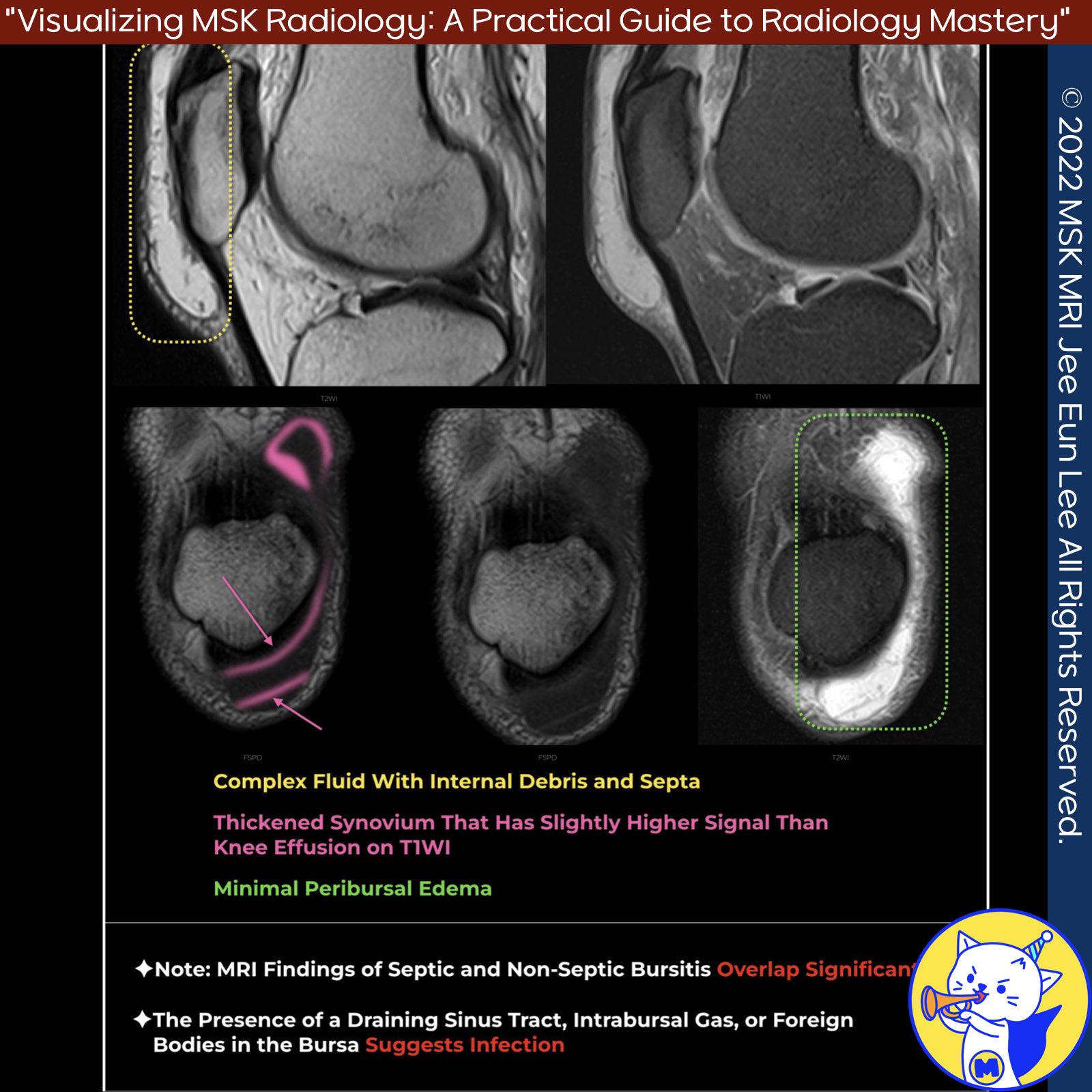
📌 Septic Bursitis
✅ Overview and Causes
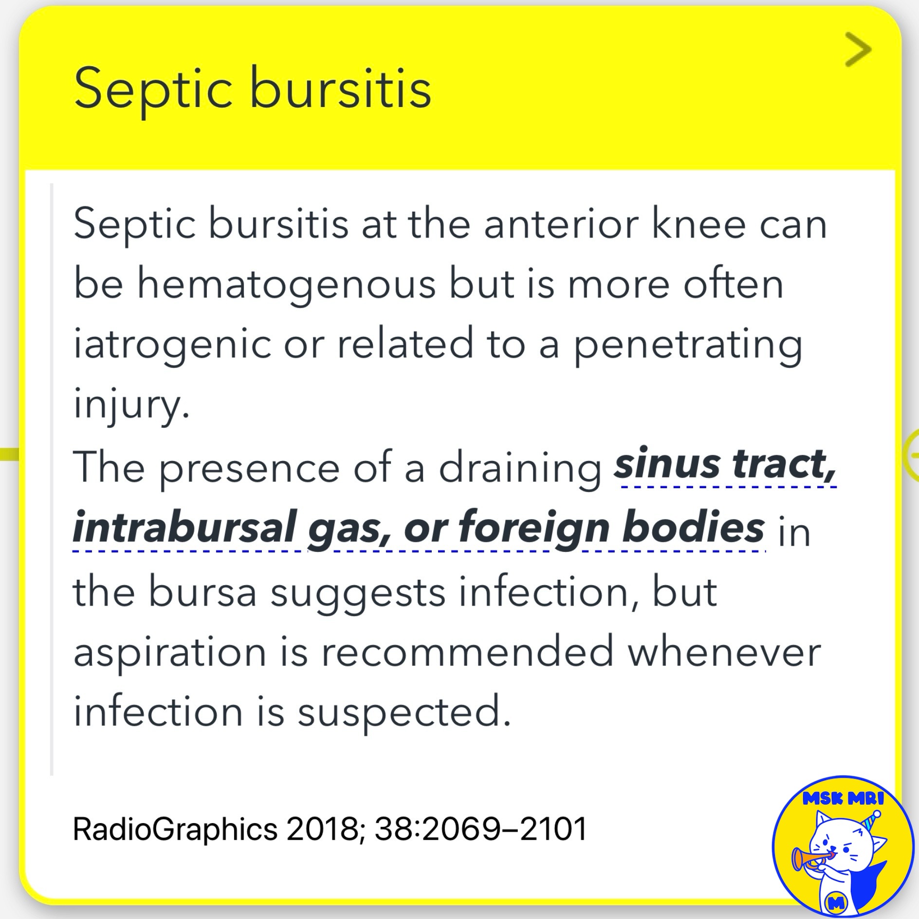
- Septic bursitis at the anterior knee can be hematogenous but is more often iatrogenic or related to a penetrating injury.
- The presence of a draining sinus tract, intrabursal gas, or foreign bodies in the bursa suggests infection, but aspiration is recommended whenever infection is suspected .
✅ Imaging Findings
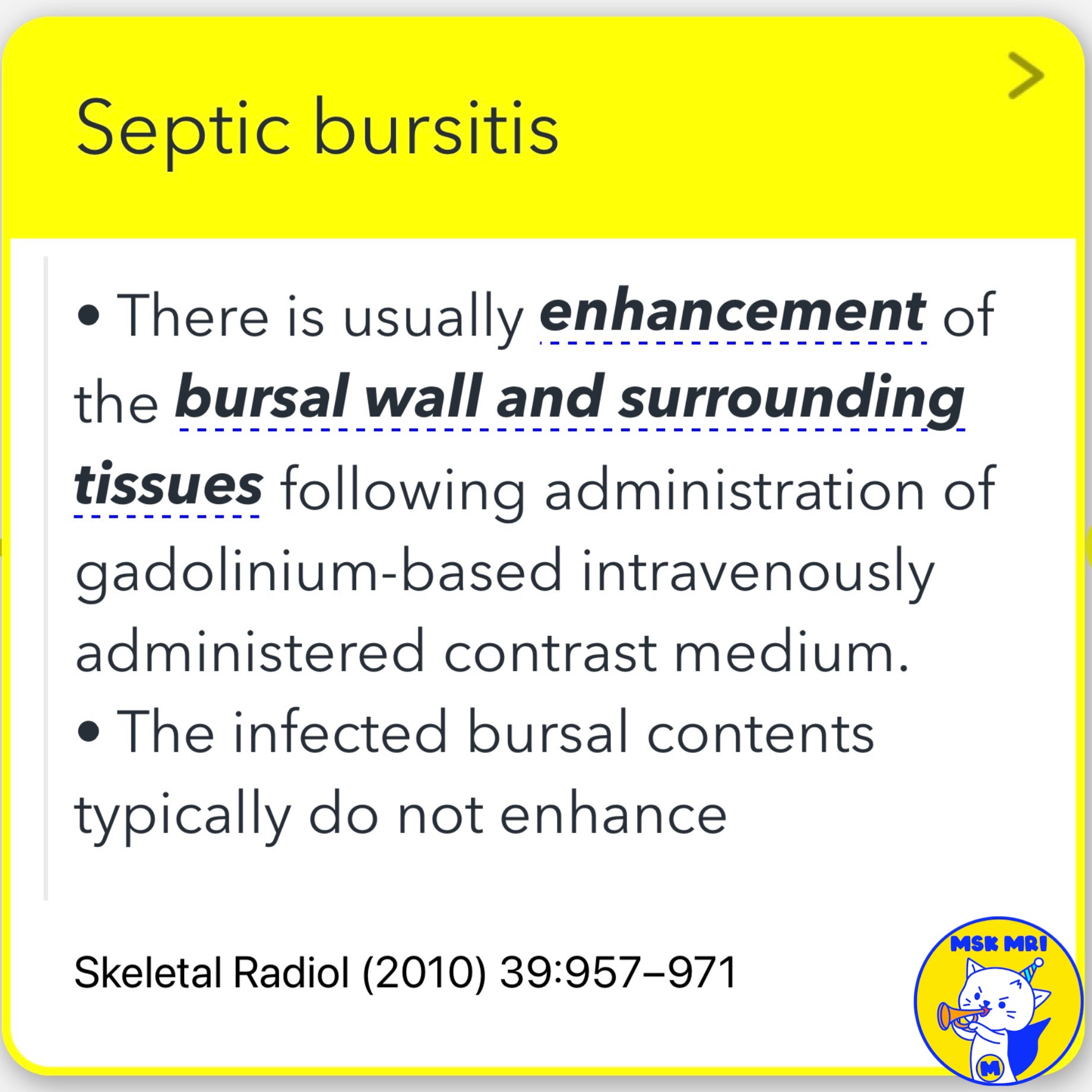
- There is usually enhancement of the bursal wall and surrounding tissues following administration of gadolinium-based intravenously administered contrast medium.
- However, the infected bursal contents typically do not enhance .
- If there is enhancement of the overlying skin, then accompanying cellulitis is invariably present. Unfortunately, there is a considerable overlap between the imaging findings of septic and nonseptic bursitis .
📌 Chronic Bursitis
- Chronic bursitis sometimes results in dramatic bursal enlargement, producing a large mass that extends well beyond the patellar borders, associated with wall thickening, loculation, and internal debris.
- This appearance is difficult to distinguish from bursal infection, as both can contain heterogeneous debris and have thickened walls that enhance after contrast material administration .
References
- RadioGraphics 2018; 38:2069–2101
- Skeletal Radiol (2010) 39:957–971
- RadioGraphics 2016; 36:1888–1910
"Visualizing MSK Radiology: A Practical Guide to Radiology Mastery"
© 2022 MSK MRI Jee Eun Lee All Rights Reserved.
No unauthorized reproduction, redistribution, or use for AI training.
#SepticBursitis, #ChronicBursitis, #Radiology, #MRI, #MusculoskeletalImaging, #Infection, #KneeInjury, #MedicalImaging, #BursalDisease, #GadoliniumContrast
'✅ Knee MRI Mastery > Chap 4BCD. Anterior knee' 카테고리의 다른 글
| (Fig 4-D.05) Deep Infrapatellar Bursitis (0) | 2024.06.23 |
|---|---|
| (Fig 4-D.04) Morel-Lavallée Lesion (0) | 2024.06.23 |
| (Fig 4-D.02) Superficial Infrapatellar Bursitis with Intrabursal Hemorrhage (0) | 2024.06.22 |
| (Fig 4-D.01) Superficial Prepatellar Bursitis in Gout (0) | 2024.06.22 |
| (Fig 4-C.14) Medial Synovial Fold of the PCL (0) | 2024.06.21 |


