==============================================
⬇️✨⬇️🎉⬇️🔥⬇️📚⬇️
Click the link to purchase on Amazon 🎉📚
==============================================
🎥 Check Out All Videos at Once! 📺
👉 Visit Visualizing MSK Blog to explore a wide range of videos! 🩻
https://visualizingmsk.blogspot.com/?view=magazine
📚 You can also find them on MSK MRI Blog and Naver Blog! 📖
https://www.instagram.com/msk_mri/
Click now to stay updated with the latest content! 🔍✨
==============================================
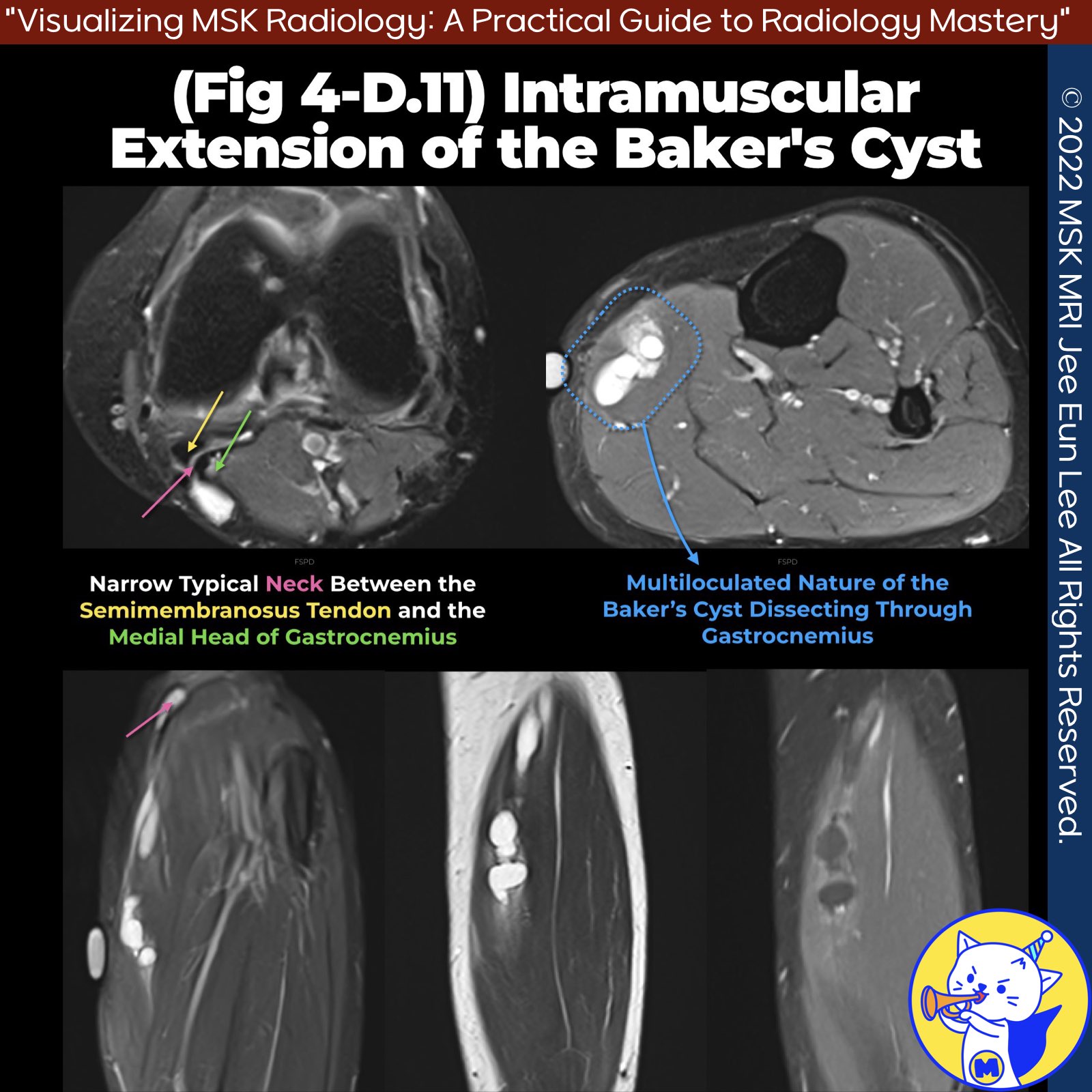
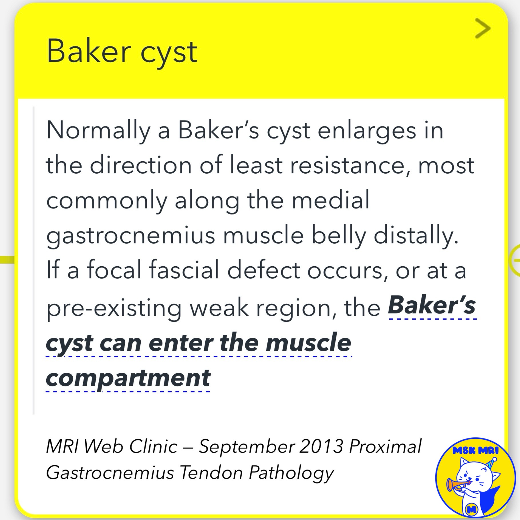
📌 Intramuscular Extension of Baker's Cyst
- A Baker’s cyst normally enlarges in the direction of least resistance, most commonly along the medial gastrocnemius muscle belly distally.
- If a focal fascial defect occurs, or at a pre-existing weak region, the Baker’s cyst can enter the muscle compartment.
✅ Dissecting Baker’s Cyst Presentation
- A dissecting Baker’s cyst may present with tender fullness at the posteromedial aspect of the knee just below the joint.
- These lesions represent rare instances where a Baker’s cyst enters through the muscle fascia and is often outside and within a muscle compartment.
Reference
- MRI Web Clinic — September 2013 Proximal Gastrocnemius Tendon Pathology
"Visualizing MSK Radiology: A Practical Guide to Radiology Mastery"
© 2022 MSK MRI Jee Eun Lee All Rights Reserved.
No unauthorized reproduction, redistribution, or use for AI training.
#BakersCyst, #IntramuscularExtension, #MusculoskeletalRadiology, #Gastrocnemius, #KneePain, #FascialDefect, #CystDissection, #KneeJoint, #RadiologyFindings, #OrthopedicRadiology


'✅ Knee MRI Mastery > Chap 4BCD. Anterior knee' 카테고리의 다른 글
| (Fig 4-D.13) Anatomical Knee Recesses: Part 1 (0) | 2024.06.25 |
|---|---|
| (Fig 4-D.12) Gastrocnemius Ganglia (0) | 2024.06.25 |
| (Fig 4-D.10) Proximal and Distal Popliteal Cyst Dissection and Rupture (0) | 2024.06.24 |
| (Fig 4-D.09) Ruptured Baker’s Cyst (0) | 2024.06.24 |
| (Fig 4-D.08) Unruptured Baker’s Cyst (0) | 2024.06.24 |