👉 Click the link below and request access—I’ll approve it for you shortly!
https://www.notion.so/MSKMRI-KNEE-b6cbb1e1bc4741b681ecf6a40159a531?pvs=4
==============================================
✨ Join the channel to enjoy the benefits! 🚀
https://www.youtube.com/channel/UC4bw7o0l2rhxn1GJZGDmT9w/join
==============================================
👉 "Click the link to purchase on Amazon 🎉📚"
[Visualizing MSK Radiology: A Practical Guide to Radiology Mastery]
https://www.amazon.com/dp/B0DJGMHMFS
==============================================
MSK MRI Jee Eun Lee
📚 Visualizing MSK Radiology: A Practical Guide to Radiology Mastery Now! 🌟 Available on Amazon, eBay, and Rain Collectibles! 💻 Ebook coming soon – stay tuned! ⏳ 🔗 https://www.amazon.com/dp/B0DJGMHMFS 🔗 https://www.ebay.com/itm/3875004193
www.youtube.com
Visualizing MSK Radiology: A Practical Guide to Radiology Mastery
www.amazon.com
📌 Ossification Variants of the Knee
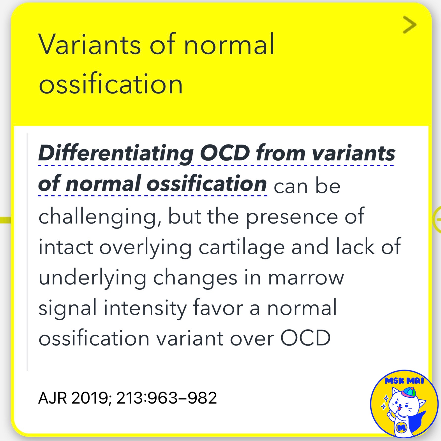
✅ Variants of Normal Ossification
- Differentiating osteochondritis dissecans (OCD) from variants of normal ossification can be challenging.
- However, the presence of intact overlying cartilage and a lack of underlying changes in marrow signal intensity favor a normal ossification variant over OCD.
- The absence of bone marrow edema, morphology, location of the lesion, and the patient's age are crucial in distinguishing a developmental variant of ossification from OCD.
- A continuous secondary physis overlying a region of osseous irregularity, with a lack of physeal disruption and adjacent subchondral bone marrow edema pattern, suggests a variation in normal development.
✅ Types of Ossification Variants of the Knee
First Case: Puzzle piece completely filled with bone.
- Characteristics: Continuous secondary physis overlying the region of osseous irregularity, lack of adjacent subchondral bone marrow edema pattern.
Second Case: Spiculated configuration with an irregular subchondral bone plate.
- Characteristics: Irregular and spiculated ossification.
Third Case: Additional ossification centers located within the unossified physeal cartilage.
- Characteristics: Extra ossification centers located within the non-ossified physeal cartilage, separated from the ossified portion of the femoral condyle.
References
- AJR 2019; 213:963–982
- RadioGraphics 2018; 38:1478–1495
"Visualizing MSK Radiology: A Practical Guide to Radiology Mastery"
© 2022 MSK MRI Jee Eun Lee All Rights Reserved.
No unauthorized reproduction, redistribution, or use for AI training.
#KneeOssification, #Radiology, #Musculoskeletal, #OssificationVariants, #OCD, #MedicalImaging, #BoneHealth, #MRI, #Orthopedics, #DevelopmentalVariant


