👉 Click the link below and request access—I’ll approve it for you shortly!
https://www.notion.so/MSKMRI-KNEE-b6cbb1e1bc4741b681ecf6a40159a531?pvs=4
==============================================
✨ Join the channel to enjoy the benefits! 🚀
https://www.youtube.com/channel/UC4bw7o0l2rhxn1GJZGDmT9w/join
==============================================
👉 "Click the link to purchase on Amazon 🎉📚"
[Visualizing MSK Radiology: A Practical Guide to Radiology Mastery]
https://www.amazon.com/dp/B0DJGMHMFS
==============================================
MSK MRI Jee Eun Lee
📚 Visualizing MSK Radiology: A Practical Guide to Radiology Mastery Now! 🌟 Available on Amazon, eBay, and Rain Collectibles! 💻 Ebook coming soon – stay tuned! ⏳ 🔗 https://www.amazon.com/dp/B0DJGMHMFS 🔗 https://www.ebay.com/itm/3875004193
www.youtube.com
Visualizing MSK Radiology: A Practical Guide to Radiology Mastery
www.amazon.com
📌 Subchondral Insufficiency Fracture (SIF)
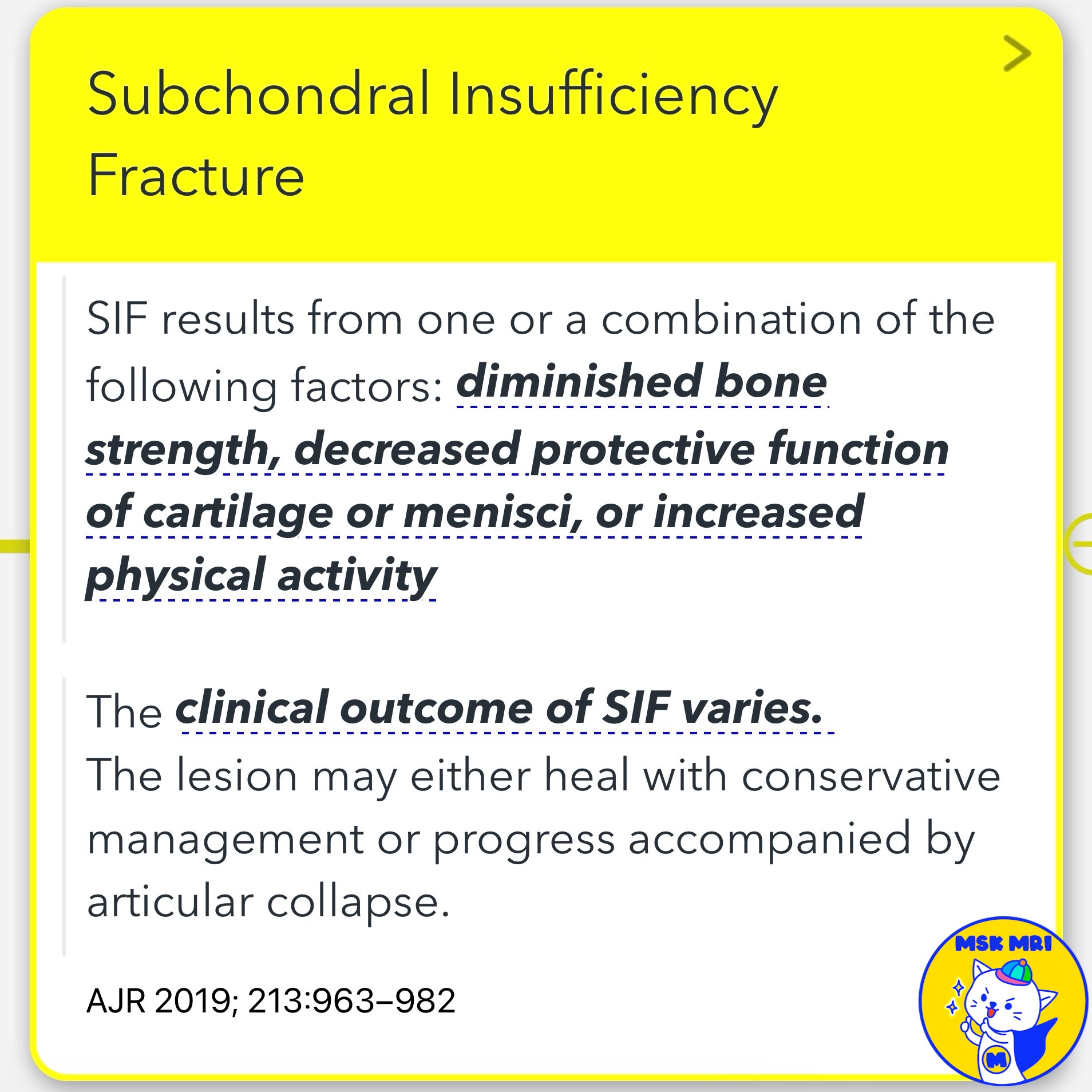
- Subchondral insufficiency fracture (SIF) results from diminished bone strength, decreased protective function of cartilage or menisci, or increased physical activity.
- The clinical outcome of SIF varies. The lesion may either heal with conservative management or progress accompanied by articular collapse.
- SIFs typically occur along the central weight-bearing aspect of the femoral condyle (60%–90%), but they may also involve the central tibial plateau and the periphery of the articular surface.
✅ MRI Findings in SIF
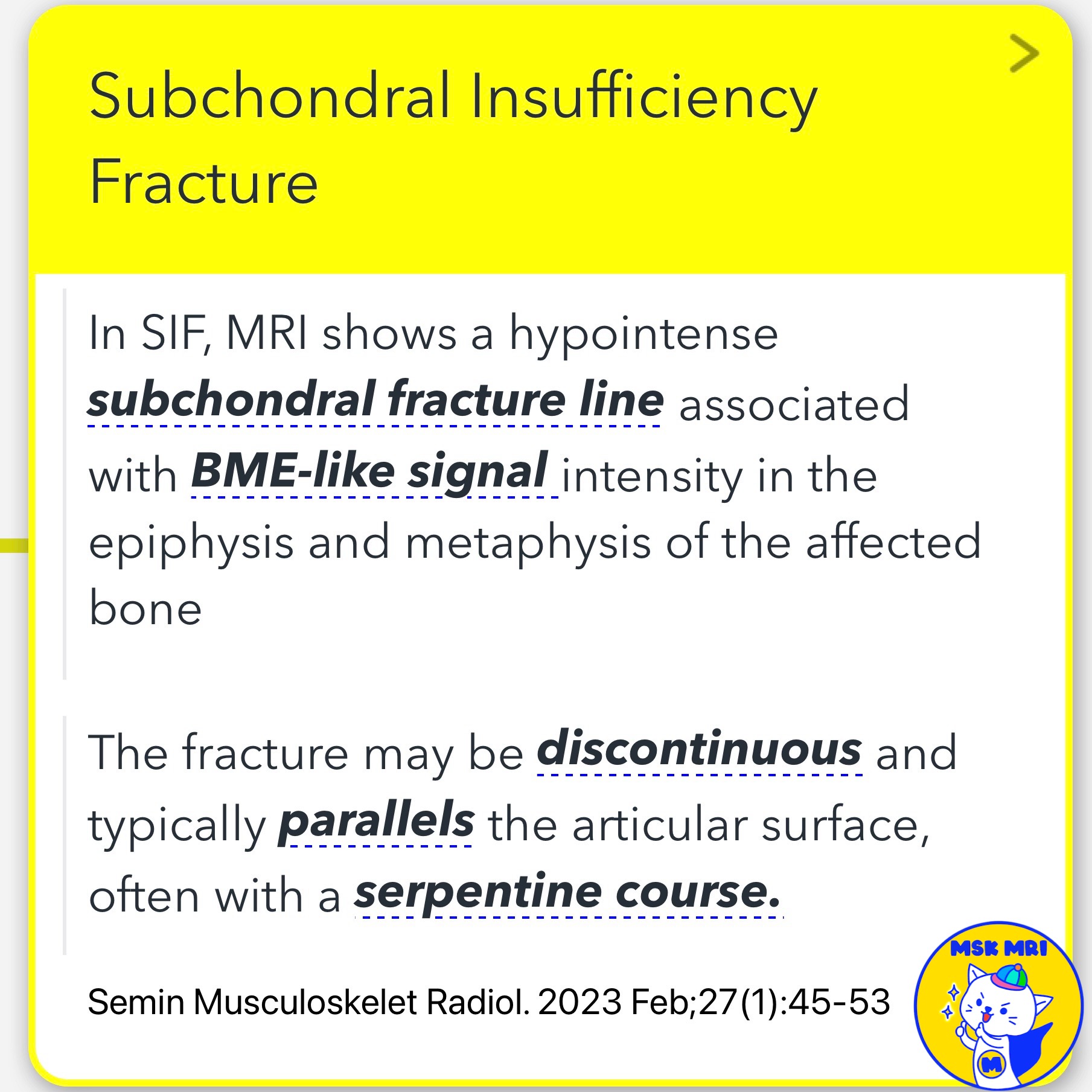
- MRI shows a hypointense subchondral fracture line with BME-like signal intensity in the epiphysis and metaphysis of the affected bone.
- The fracture may be discontinuous and typically parallels the articular surface with a serpentine course.
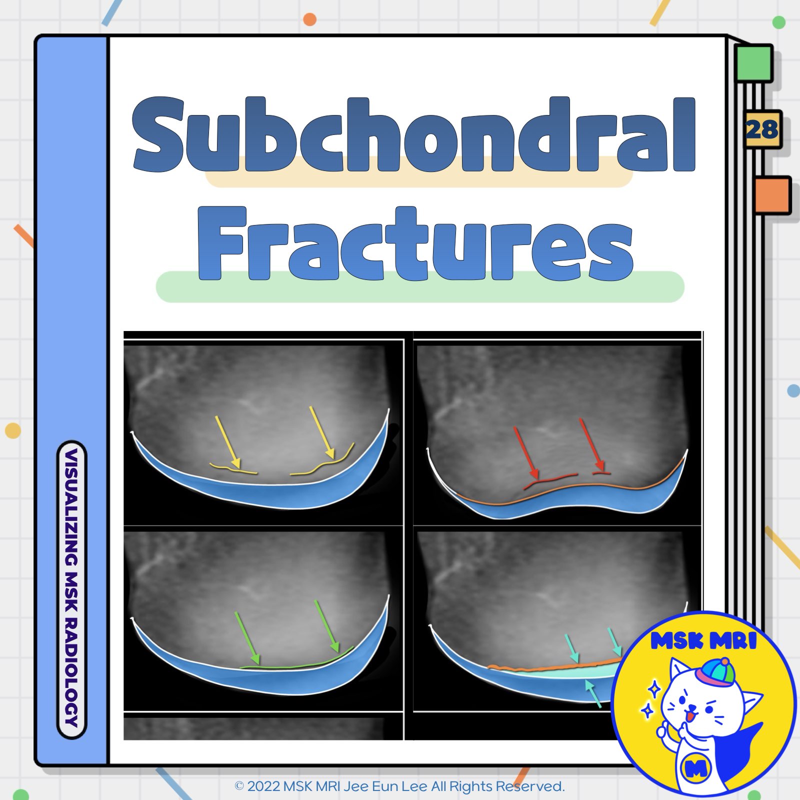
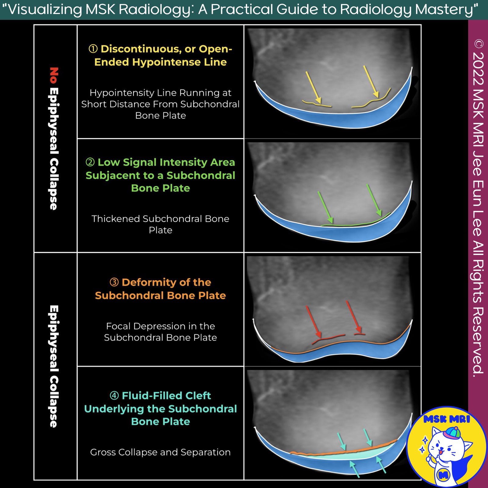
- SIF can be categorized based on the presence or absence of epiphyseal collapse:
★ Without Epiphyseal Collapse:
1. Discontinuous or open-ended hypointense fracture line near the subchondral bone plate.
2. Low signal intensity area under the subchondral bone plate with a thickened subchondral bone plate.
★ With Epiphyseal Collapse:
3. Deformity and focal depression in the subchondral bone plate.
4. Fluid-filled cleft under the subchondral bone plate indicating gross collapse and separation.
📌 Spontaneous Osteonecrosis of the Knee (SONK), (misnomer)
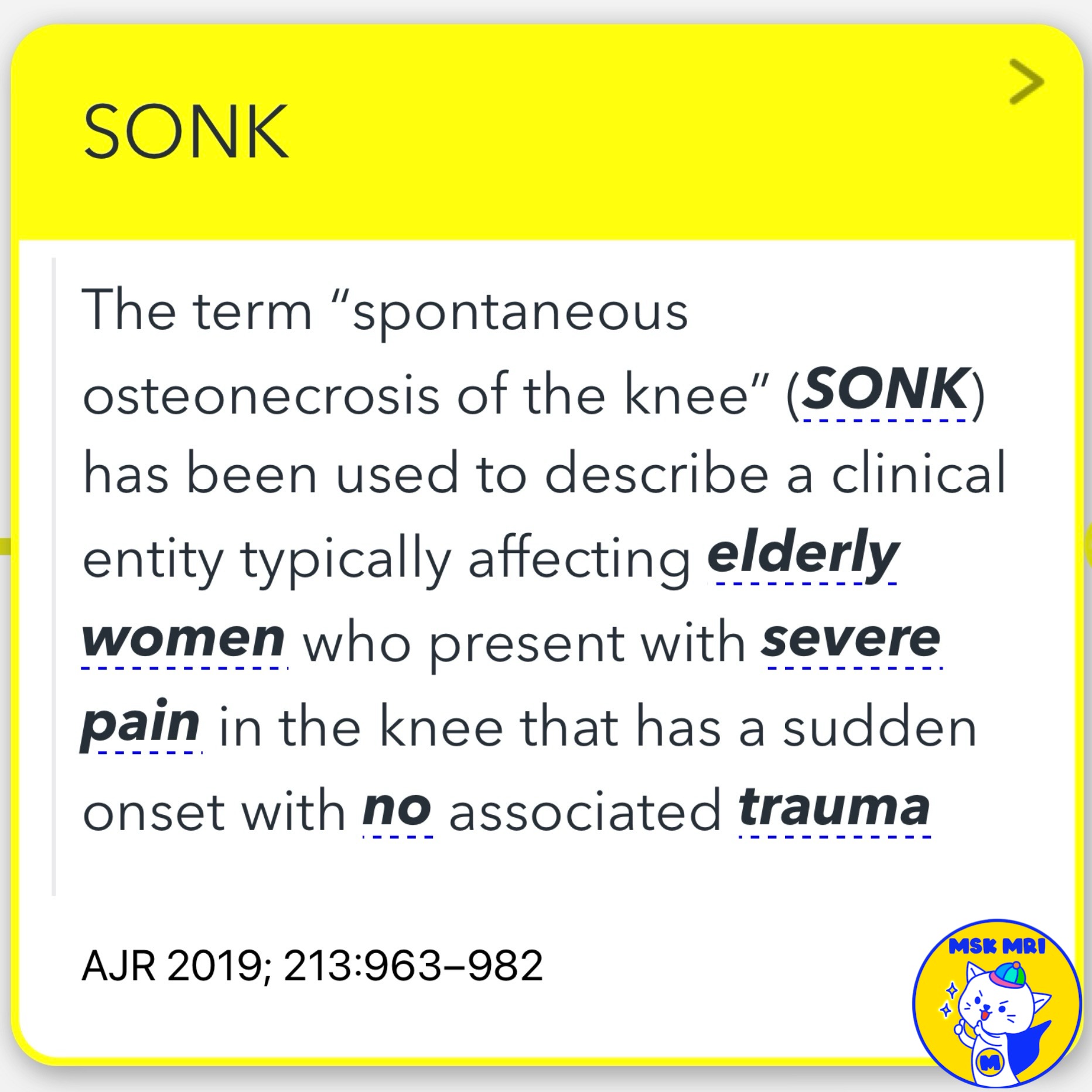
- “Spontaneous osteonecrosis of the knee” (SONK) describes a condition typically affecting elderly women with sudden, severe knee pain and no associated trauma.
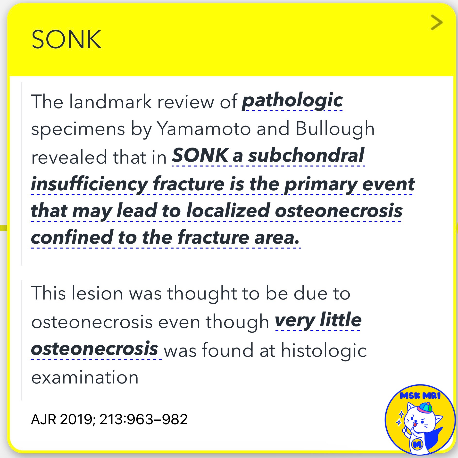
- Yamamoto and Bullough's review revealed that in SONK, a subchondral insufficiency fracture is the primary event leading to localized osteonecrosis confined to the fracture area. Histologic examination showed very little osteonecrosis.
✅ Terminology
SONK is now considered a misnomer, representing a SIF of the knee that has progressed to subchondral collapse and secondary osteonecrosis. Some authors suggest the term SONK for lesions that will not heal, proposing "irreversible SIF" or "pseudoarthrosis" as more appropriate terms.
References
- AJR 2019; 213:963–982
- Semin Musculoskelet Radiol. 2023 Feb;27(1):45-53
- Semin Musculoskelet Radiol. 2023 Feb;27(1):103-113
"Visualizing MSK Radiology: A Practical Guide to Radiology Mastery"
© 2022 MSK MRI Jee Eun Lee All Rights Reserved.
No unauthorized reproduction, redistribution, or use for AI training.
#SubchondralInsufficiencyFracture, #SIF, #MRI, #BoneMarrowEdema, #EpiphysealCollapse, #FemoralCondyle, #SubchondralBonePlate, #SONK, #Osteonecrosis, #Pseudoarthrosis

