👉 Click the link below and request access—I’ll approve it for you shortly!
https://www.notion.so/MSKMRI-KNEE-b6cbb1e1bc4741b681ecf6a40159a531?pvs=4
==============================================
✨ Join the channel to enjoy the benefits! 🚀
https://www.youtube.com/channel/UC4bw7o0l2rhxn1GJZGDmT9w/join
==============================================
👉 "Click the link to purchase on Amazon 🎉📚"
[Visualizing MSK Radiology: A Practical Guide to Radiology Mastery]
https://www.amazon.com/dp/B0DJGMHMFS
==============================================
MSK MRI Jee Eun Lee
📚 Visualizing MSK Radiology: A Practical Guide to Radiology Mastery Now! 🌟 Available on Amazon, eBay, and Rain Collectibles! 💻 Ebook coming soon – stay tuned! ⏳ 🔗 https://www.amazon.com/dp/B0DJGMHMFS 🔗 https://www.ebay.com/itm/3875004193
www.youtube.com
Visualizing MSK Radiology: A Practical Guide to Radiology Mastery
www.amazon.com
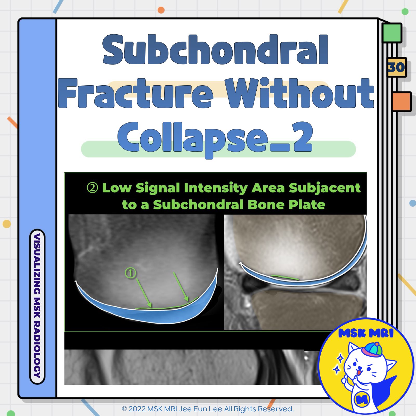
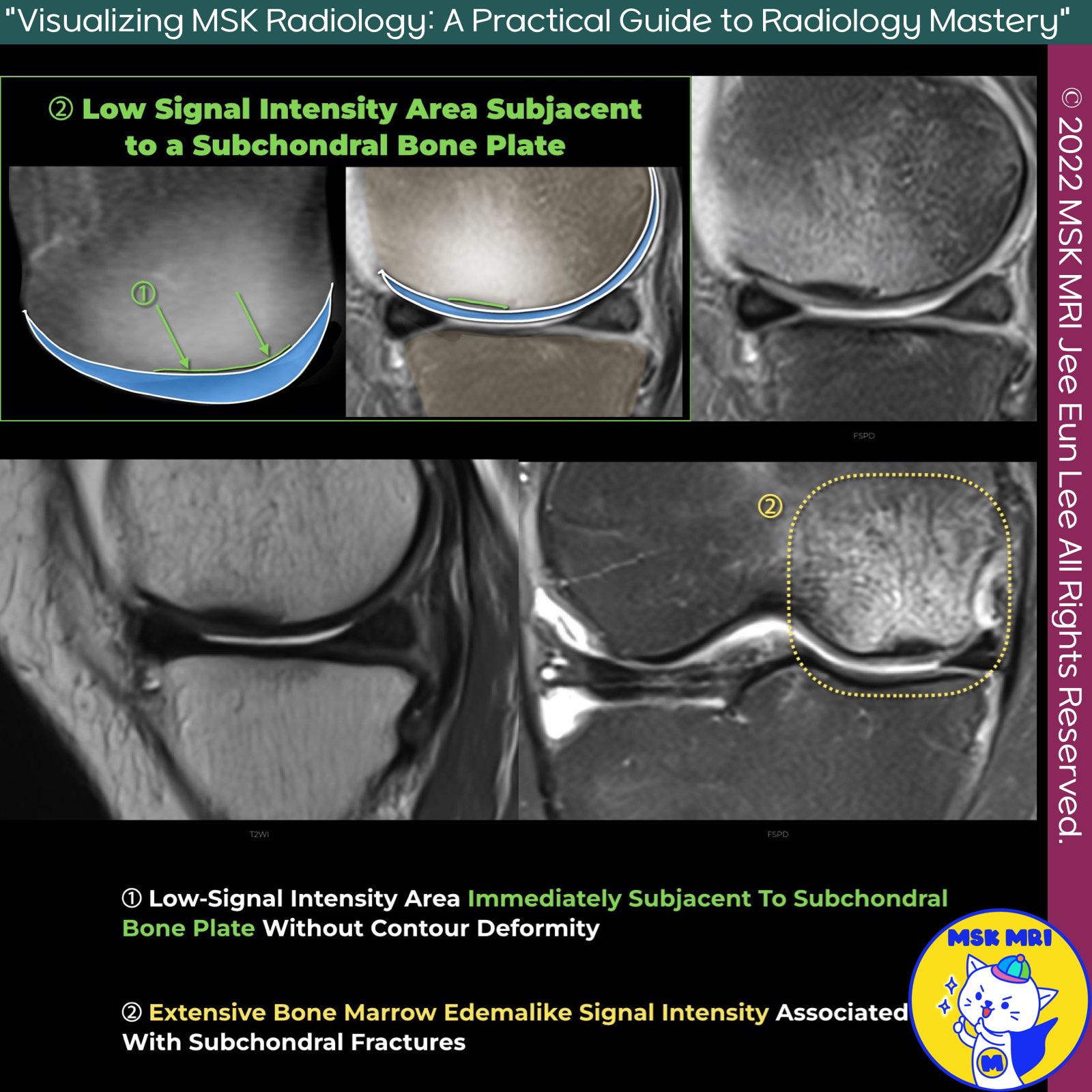
✅ Low Signal Intensity Area Subjacent to a Subchondral Bone Plate without Epiphyseal Collapse
1. Low-Signal Intensity Area Subjacent to Subchondral Bone Plate Without Contour Deformity
- Description: This finding appears as a low signal intensity fracture line running very close to the subchondral bone plate, giving the appearance of a thickened subchondral bone plate.
- Reason: This occurs due to a combination of a fracture with callus and granulation tissue and secondary osteonecrosis between the fracture line and the articular surface.
2. Extensive Bone Marrow Edemalike Signal Intensity Associated with Subchondral Fractures
- Description: This finding presents extensive bone marrow edema-like signal intensity related to subchondral fractures.
Additional Considerations
- Radiologists should note that the subchondral area of low signal intensity often integrates with the subchondral plate and should not be mistaken for a thickened subchondral plate.
- A dedicated MRI with a small Field of View (FOV) is recommended to evaluate individual morphologic findings in SIF, such as fracture lines, subchondral hypointense areas, subtle contour changes, cartilage loss, and the presence of collapse.
References
- RadioGraphics 2018; 38:1478–1495
- AJR 2019; 213:963–982
- Japanese Journal of Radiology (2022) 40:443–457
"Visualizing MSK Radiology: A Practical Guide to Radiology Mastery"
© 2022 MSK MRI Jee Eun Lee All Rights Reserved.
No unauthorized reproduction, redistribution, or use for AI training.
#MRI #Radiology #SubchondralInsufficiencyFracture #SIF #EpiphysealCollapse #BoneMarrowEdema #SubchondralFracture #Radiologist #MedicalImaging #OrthopedicRadiology


