👉 Click the link below and request access—I’ll approve it for you shortly!
https://www.notion.so/MSKMRI-KNEE-b6cbb1e1bc4741b681ecf6a40159a531?pvs=4
==============================================
✨ Join the channel to enjoy the benefits! 🚀
https://www.youtube.com/channel/UC4bw7o0l2rhxn1GJZGDmT9w/join
==============================================
👉 "Click the link to purchase on Amazon 🎉📚"
[Visualizing MSK Radiology: A Practical Guide to Radiology Mastery]
https://www.amazon.com/dp/B0DJGMHMFS
==============================================
MSK MRI Jee Eun Lee
📚 Visualizing MSK Radiology: A Practical Guide to Radiology Mastery Now! 🌟 Available on Amazon, eBay, and Rain Collectibles! 💻 Ebook coming soon – stay tuned! ⏳ 🔗 https://www.amazon.com/dp/B0DJGMHMFS 🔗 https://www.ebay.com/itm/3875004193
www.youtube.com
Visualizing MSK Radiology: A Practical Guide to Radiology Mastery
www.amazon.com

📌 Septic Arthritis
✅ Noncontrast MR Imaging
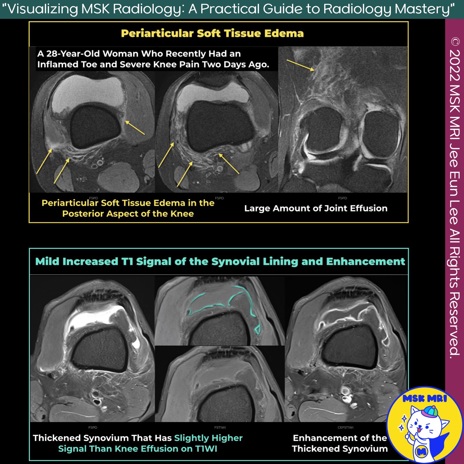
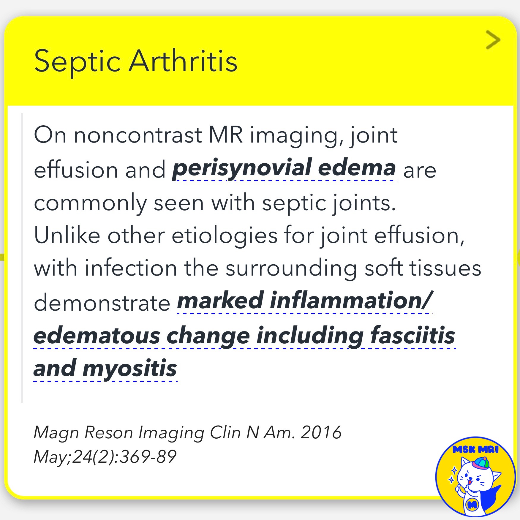
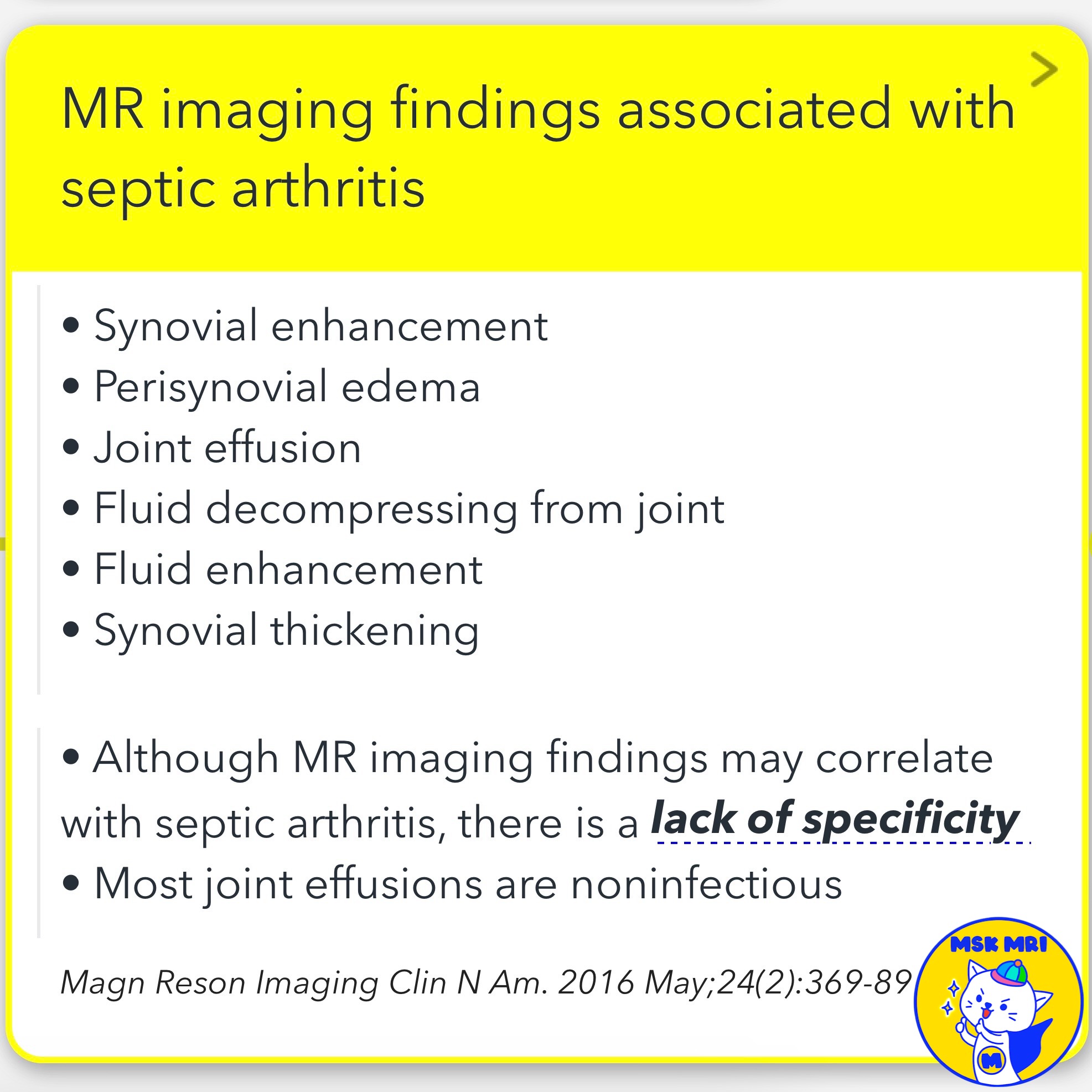
- On noncontrast MR imaging, joint effusion and perisynovial edema are commonly seen with septic joints.
- Unlike other etiologies for joint effusion, with infection, the surrounding soft tissues demonstrate marked inflammation and edematous changes, including fasciitis and myositis.
✅ Postcontrast Imaging
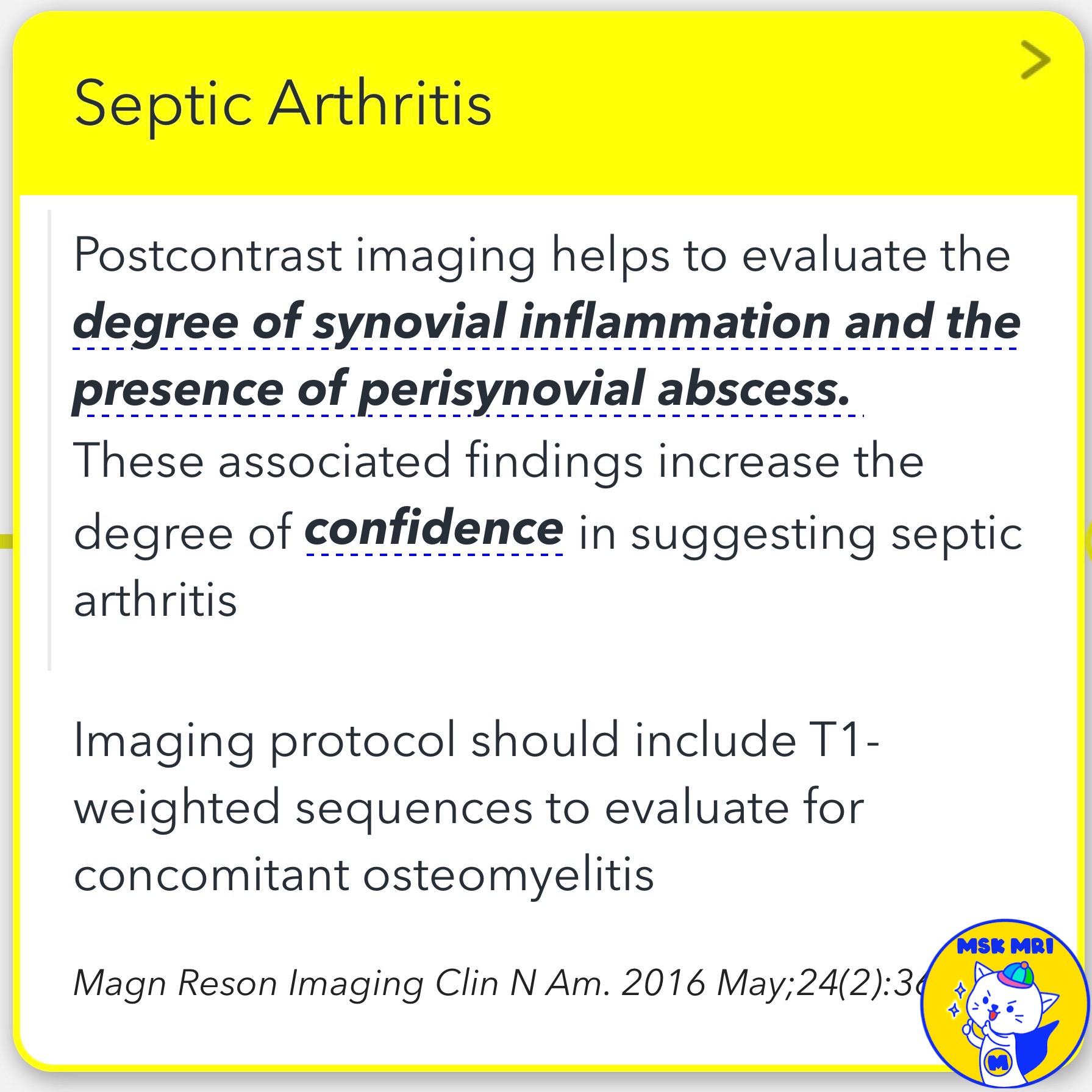
- Postcontrast imaging helps to evaluate the degree of synovial inflammation and the presence of perisynovial abscess.
- These associated findings increase the degree of confidence in suggesting septic arthritis.
- Imaging protocol should include T1-weighted sequences to evaluate for concomitant osteomyelitis.
✅ Earliest Findings of Septic Arthritis
The earliest findings in septic arthritis, including synovial inflammation and effusions, are readily demonstrated.
✅ Acute Stage of Septic Arthritis
- Bone marrow signal abnormality is also a frequent finding in the acute stage.
- At this stage, bone marrow edema usually represents inflammation.
- If contrast is administered intravenously, a marked rim enhancement of the synovium or of a thickened abscess wall (if present) can be seen.
References
- Magn Reson Imaging Clin N Am. 2016 May;24(2):369-89
- Magn Reson Imaging Clin N Am 12 (2004) 111–124
"Visualizing MSK Radiology: A Practical Guide to Radiology Mastery"
© 2022 MSK MRI Jee Eun Lee All Rights Reserved.
No unauthorized reproduction, redistribution, or use for AI training.
#SepticArthritis, #MRI, #Radiology, #JointEffusion, #SynovialInflammation, #PerisynovialEdema, #Osteomyelitis, #Fasciitis, #Myositis, #BoneMarrowEdema

'✅ Knee MRI Mastery > Chap 5E. Other' 카테고리의 다른 글
| (Fig 5-E.06) Intracapsular Locations of Tophi, Gout (0) | 2024.07.23 |
|---|---|
| (Fig 5-E.05) Rheumatoid Arthritis - Rice Bodies (1) | 2024.07.23 |
| (Fig 5-E.04) Rheumatoid Arthritis - Erosion and Synovitis (2) | 2024.07.23 |
| (Fig 5-E.03) MRI Findings of Septic Arthritis for Therapy Monitoring (0) | 2024.07.22 |
| (Fig 5-E.02) Bone Erosions in Pyogenic Arthritis (0) | 2024.07.22 |