👉 Click the link below and request access—I’ll approve it for you shortly!
https://www.notion.so/MSKMRI-KNEE-b6cbb1e1bc4741b681ecf6a40159a531?pvs=4
==============================================
✨ Join the channel to enjoy the benefits! 🚀
https://www.youtube.com/channel/UC4bw7o0l2rhxn1GJZGDmT9w/join
==============================================
👉 "Click the link to purchase on Amazon 🎉📚"
[Visualizing MSK Radiology: A Practical Guide to Radiology Mastery]
https://www.amazon.com/dp/B0DJGMHMFS
==============================================
MSK MRI Jee Eun Lee
📚 Visualizing MSK Radiology: A Practical Guide to Radiology Mastery Now! 🌟 Available on Amazon, eBay, and Rain Collectibles! 💻 Ebook coming soon – stay tuned! ⏳ 🔗 https://www.amazon.com/dp/B0DJGMHMFS 🔗 https://www.ebay.com/itm/3875004193
www.youtube.com
Visualizing MSK Radiology: A Practical Guide to Radiology Mastery
www.amazon.com

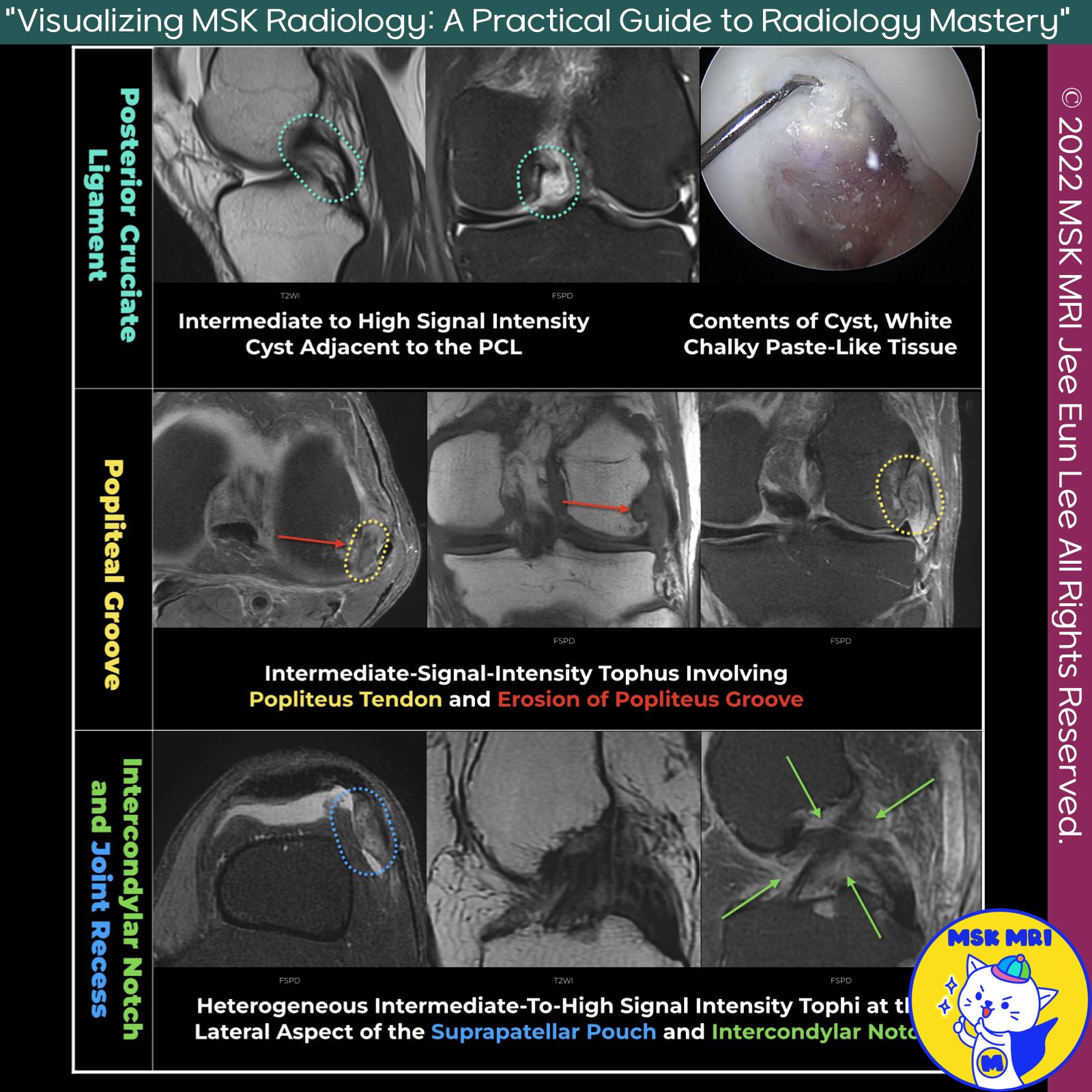
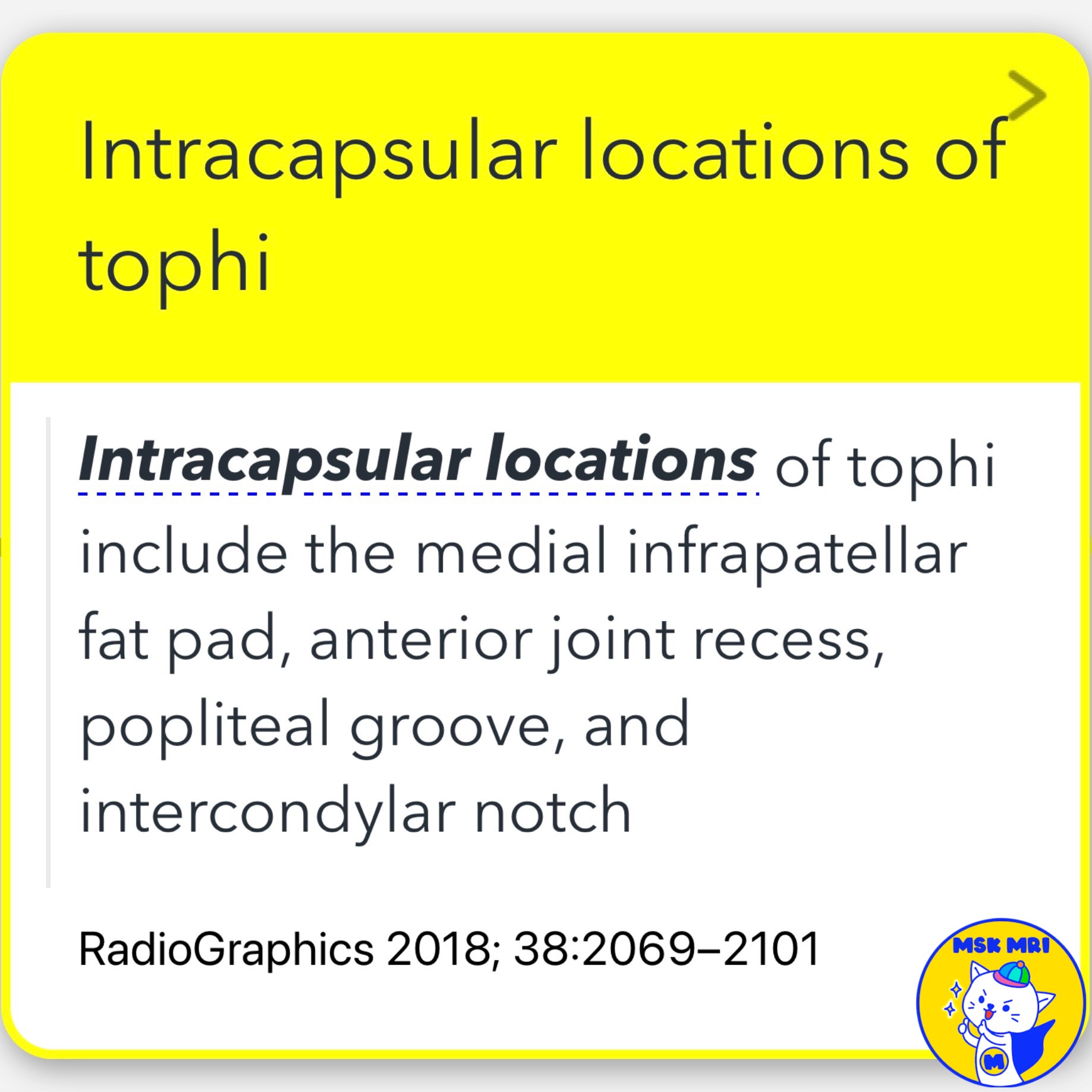
📌Intracapsular Locations of Tophi
- Tophi can be found in several intracapsular locations, including the medial infrapatellar fat pad, anterior joint recess, popliteal groove, and intercondylar notch.
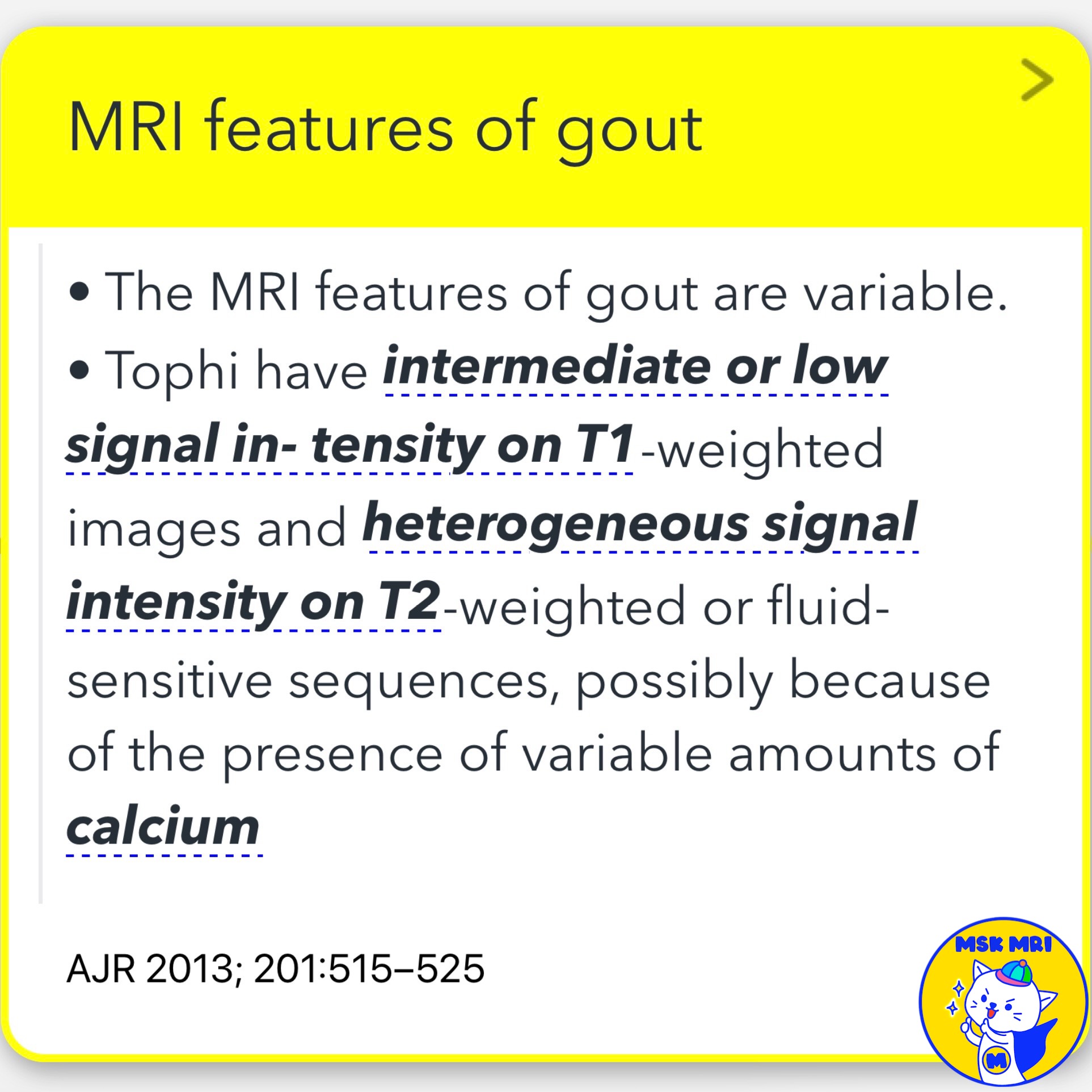
✅ MRI Features of Gout
- The MRI features of gout are variable.
- Tophi exhibit intermediate or low signal intensity on T1-weighted images and heterogeneous signal intensity on T2-weighted or fluid-sensitive sequences, possibly due to varying amounts of calcium present.
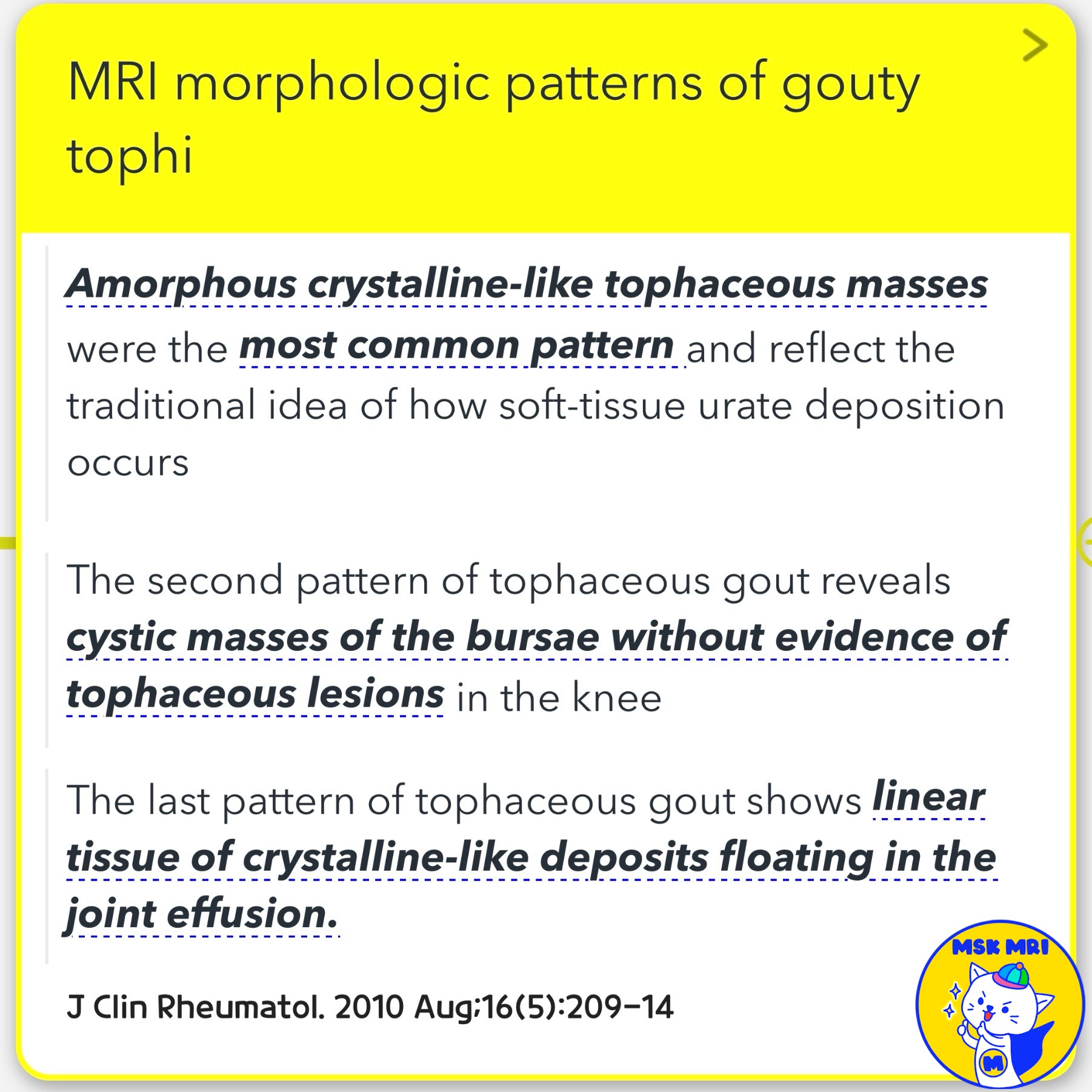
✅ MRI Morphologic Patterns of Gouty Tophi
- Amorphous Crystalline-like Tophi: The most common pattern, reflecting the traditional concept of soft-tissue urate deposition.
- Cystic Masses in Bursae: The second pattern shows cystic masses in the bursae without evidence of tophaceous lesions in the knee.
- Linear Crystalline-like Deposits: The final pattern features linear tissue of crystalline-like deposits floating in the joint effusion.
References
- RadioGraphics 2018; 38:2069–2101
- AJR 2013; 201:515–525
- J Clin Rheumatol. 2010 Aug;16(5):209-14
"Visualizing MSK Radiology: A Practical Guide to Radiology Mastery"
© 2022 MSK MRI Jee Eun Lee All Rights Reserved.
No unauthorized reproduction, redistribution, or use for AI training.
#Gout, #Tophi, #MRI, #Radiology, #InfrapatellarFatPad, #JointRecess, #PoplitealGroove, #IntercondylarNotch, #TophaceousGout, #CrystallineDeposits
'✅ Knee MRI Mastery > Chap 5E. Other' 카테고리의 다른 글
| (Fig 5-E.08) Acute Gouty Arthritis vs. Septic Arthritis (0) | 2024.07.23 |
|---|---|
| (Fig 5-E.07) Extracapsular Locations of Tophi (0) | 2024.07.23 |
| (Fig 5-E.05) Rheumatoid Arthritis - Rice Bodies (1) | 2024.07.23 |
| (Fig 5-E.04) Rheumatoid Arthritis - Erosion and Synovitis (2) | 2024.07.23 |
| (Fig 5-E.03) MRI Findings of Septic Arthritis for Therapy Monitoring (0) | 2024.07.22 |