https://youtube.com/shorts/4iPGfQrwxwY

Coalition by MSKMRI JEE EUN LEE.pdf
6.67MB
The Harris heel (axial) view is a specialized radiograph designed to evaluate the subtalar joint, particularly for detecting talocalcaneal coalitions.
Normal Harris view
- Structures: calcaneal body, middle facet, posterior facet (anterior facet obscured by talar head).
- Sustentaculum tali projects clearly from the medial calcaneus.
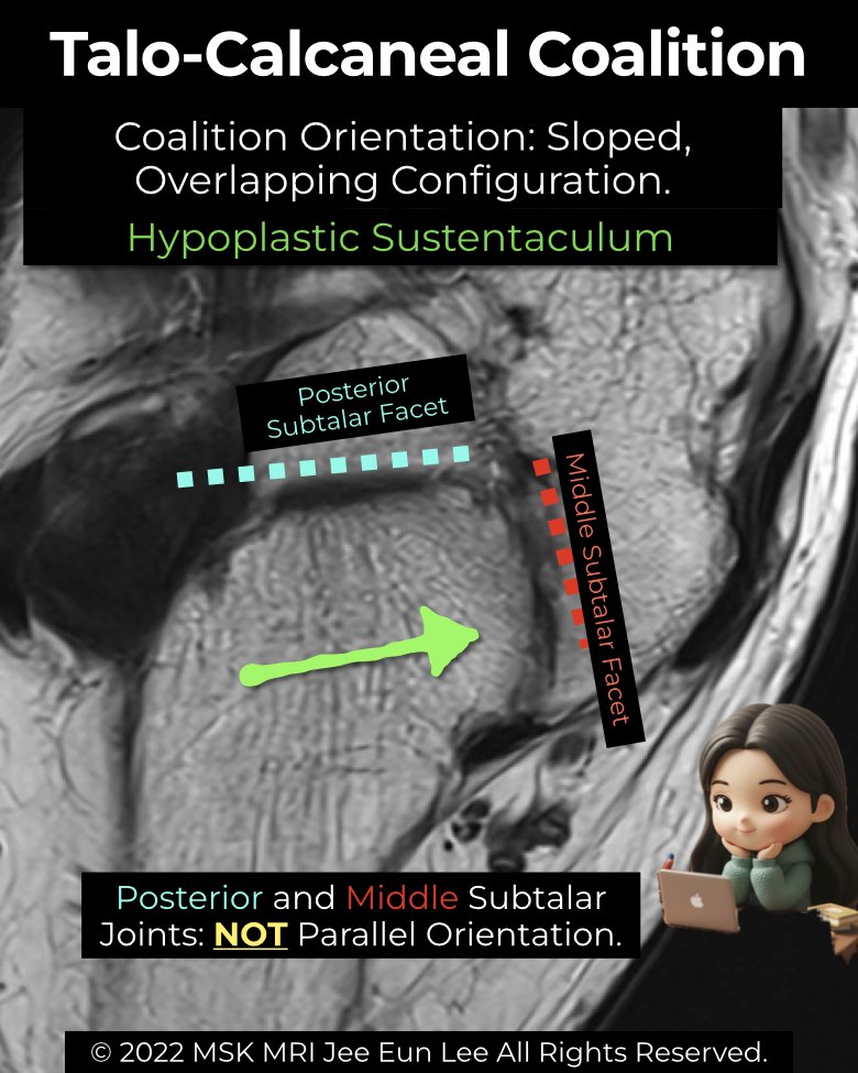
- Key feature: parallel orientation of middle and posterior subtalar facets (<20°).
Abnormal Harris view (coalition)
- Non-osseous (fibrous/cartilaginous):
- Osseous (synostosis):
Radiology perspective
- A normal Harris view reassures with parallel middle and posterior facets.
- Any loss of parallelism, irregular cleft, or bony bridge strongly suggests coalition.
- Still valuable as a screening tool, though CT and MRI remain gold standards for full characterization.
#Radiology, #MSKMRI, #HarrisHeelView, #SubtalarJoint, #CoalitionImaging, #FootXray, #RadiologyEducation, #OrthopedicImaging, #MSKImaging, #RadiologistLife
Visualizing MSK Radiology: A Practical Guide to Radiology Mastery
© 2022 MSK MRI Jee Eun Lee All Rights Reserved.
No unauthorized reproduction, redistribution, or use for AI training.