https://youtube.com/shorts/4TviAMKn8d4

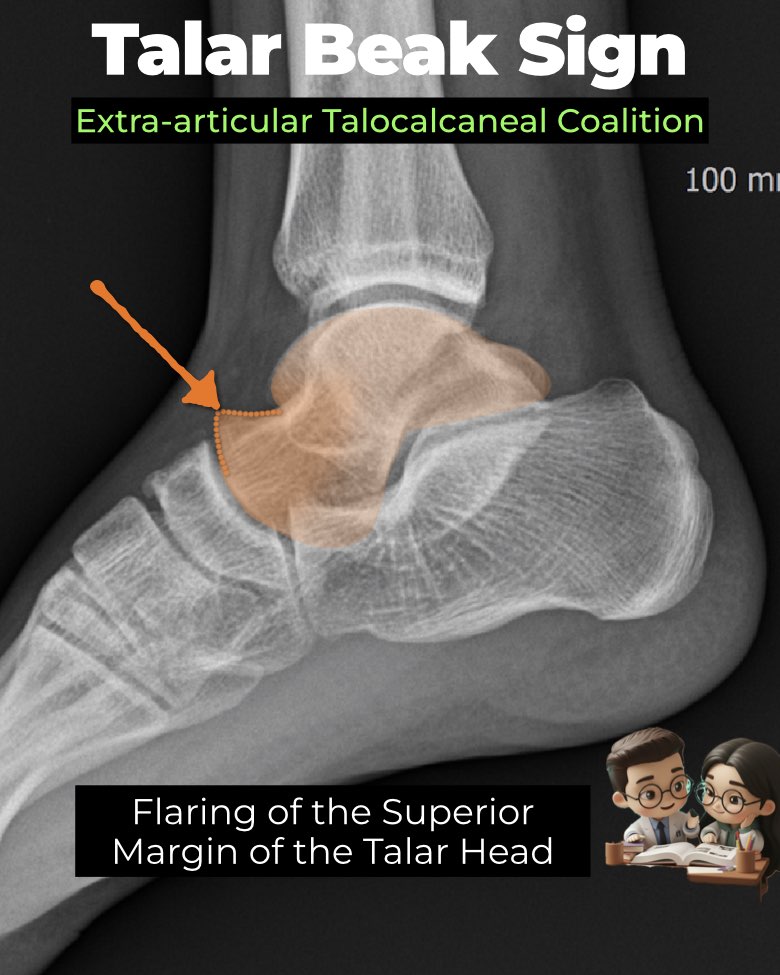
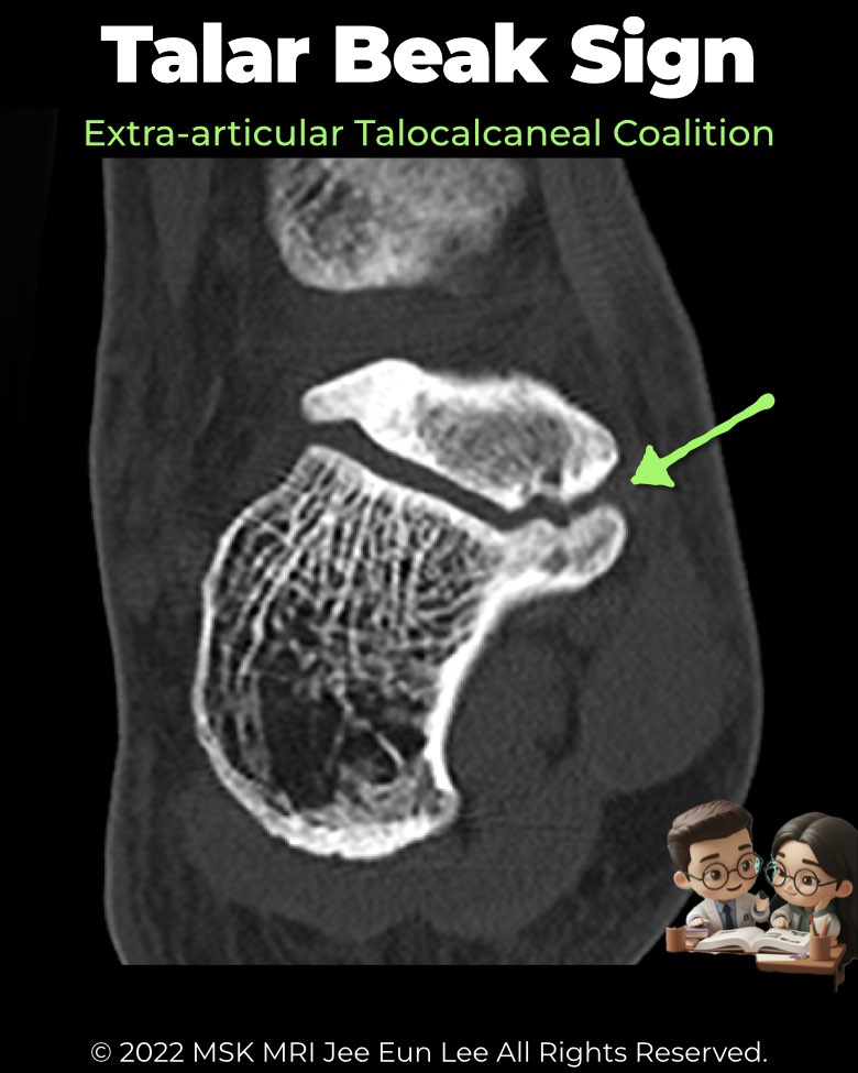
A talar beak is a classic indirect radiographic sign of tarsal coalition, best appreciated on a lateral foot or ankle radiograph.
Pathophysiology
- Coalition restricts subtalar motion → compensatory hypermobility at the talonavicular joint.
- Repetitive stress at the talonavicular ligament/capsule insertion causes periosteal stripping and traction osteophyte formation.
- Result: subperiosteal bone proliferation projecting from the dorsal talar head = the “beak.”
Imaging appearance
- Location: dorsal surface of the talar head, at the talar ridge (talonavicular ligament insertion).
- Morphology: large, triangular, and sloping distally, flaring upward away from the navicular.
- Differentiation:
Diagnostic utility
- Associated most often with talocalcaneal coalitions, but can also occur in calcaneonavicular coalitions.
- Sensitivity: ~48–49%; Specificity: ~91%.
- Absence does not rule out coalition; presence strongly suggests it.
- Not seen in cuboid–navicular coalitions, since these do not alter talonavicular biomechanics.
- No prognostic significance for surgical outcomes or degenerative risk.
Radiology perspective
A talar beak is a specific but not sensitive sign—when present, strongly consider coalition, but always confirm with CT or MRI and correlate with other secondary signs (C-sign, dysmorphic sustentaculum tali).
#Radiology, #MSKMRI, #TalarBeak, #TalocalcanealCoalition, #FootXray, #CoalitionImaging, #RadiologyEducation, #OrthopedicImaging, #MSKImaging, #RadiologistLife
Visualizing MSK Radiology: A Practical Guide to Radiology Mastery
© 2022 MSK MRI Jee Eun Lee All Rights Reserved.
No unauthorized reproduction, redistribution, or use for AI training.