https://youtube.com/shorts/xm0KH7pjsvc

Isolated posterior subtalar joint coalitions are uncommon compared with the classic middle facet type.
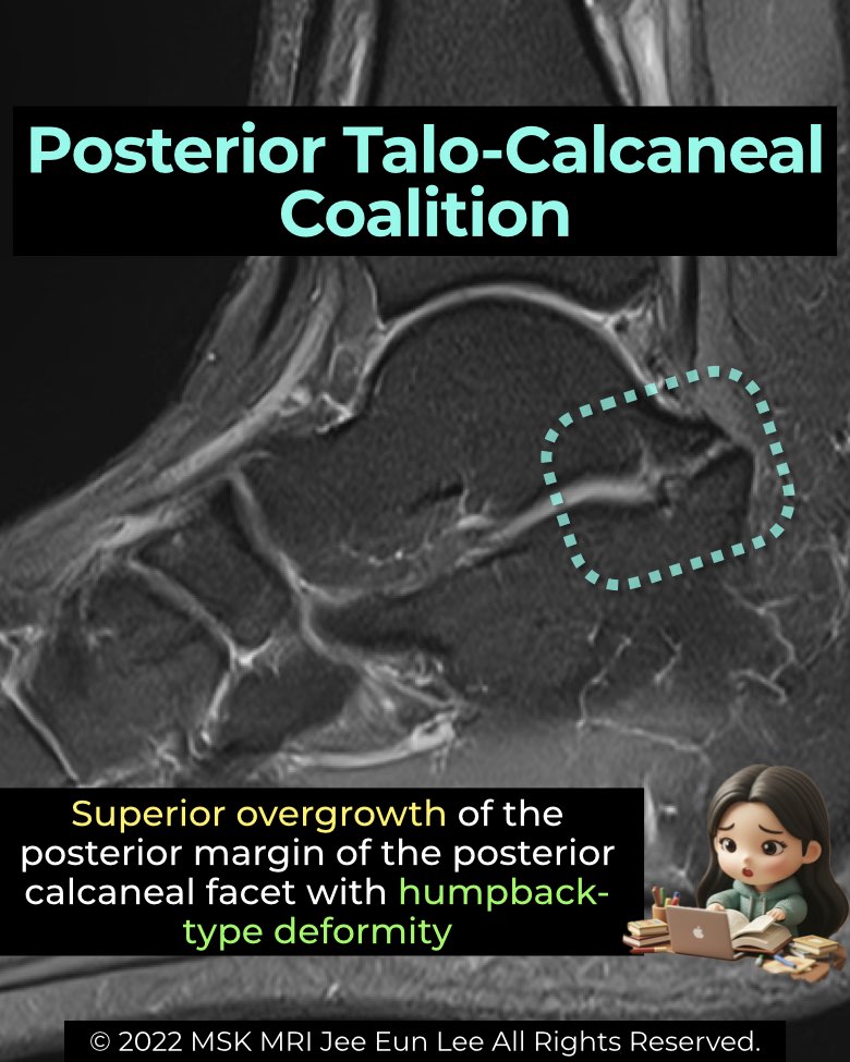
Anatomy & morphology
- Typically cartilaginous in nature.
- Involves the postero-medial portion of the posterior facet.
- May show bony overgrowth protruding into the tarsal tunnel.
- A superiorly directed spur from the posterior calcaneal facet can create a “humpback-type deformity.”
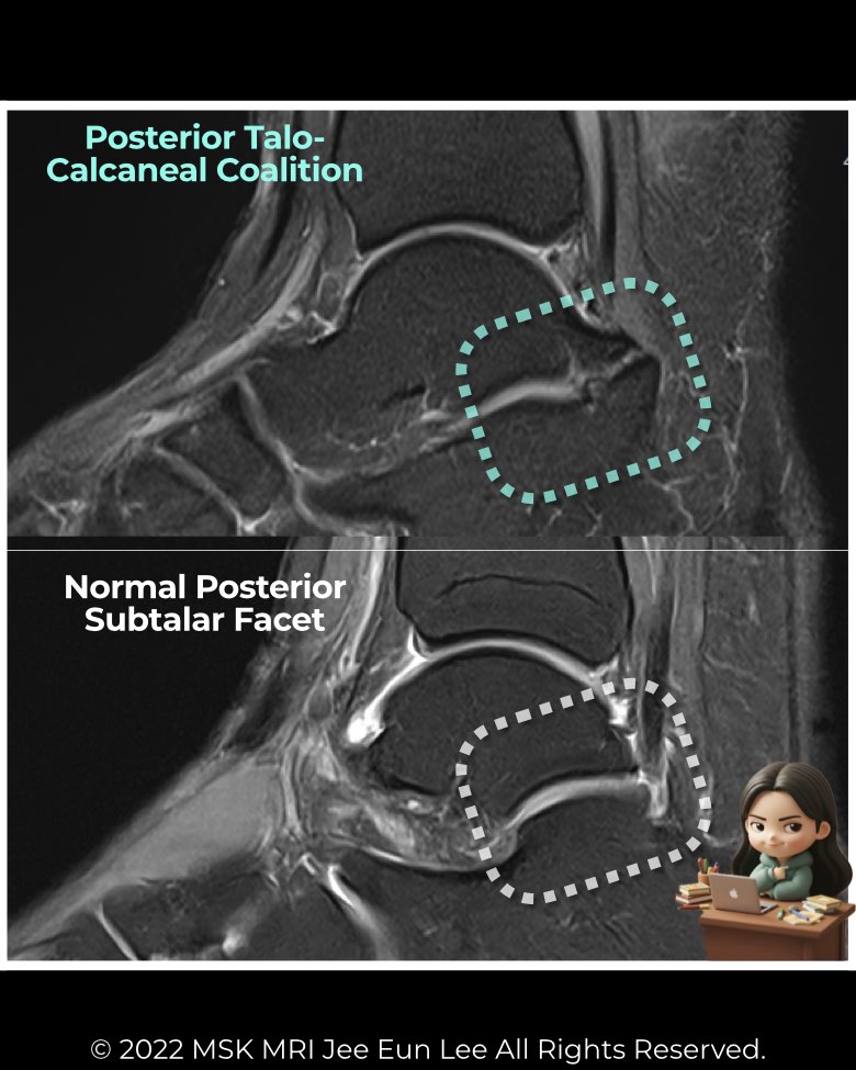
Imaging findings
- Radiographs: often subtle; indirect signs may be absent.
- CT & MRI: essential for diagnosis. MRI can depict cartilaginous bridging, marrow edema, and associated posterior facet hypertrophy.
- Look for narrowing or irregularity of the posterior facet and abnormal contour projecting medially into the tarsal tunnel.
Clinical relevance
- Symptoms: hindfoot pain, stiffness, or tarsal tunnel syndrome if overgrowth encroaches on the neurovascular bundle.
- Surgical outcomes: patients with isolated posterior facet coalitions (P-type) show better postoperative results—higher functional scores and less pain—than those with combined middle–posterior (MP-type) coalitions.
Recognizing this rare type is key, as differentiating isolated posterior involvement from combined lesions has significant implications for treatment planning and prognosis.
#Radiology, #MSKMRI, #SubtalarJoint, #PosteriorFacet, #CoalitionImaging, #FootMRI, #OrthopedicImaging, #RadiologyEducation, #MSKImaging, #RadiologistLife
Visualizing MSK Radiology: A Practical Guide to Radiology Mastery
© 2022 MSK MRI Jee Eun Lee All Rights Reserved.
No unauthorized reproduction, redistribution, or use for AI training.