https://youtube.com/shorts/cQMfJ8hTnhU

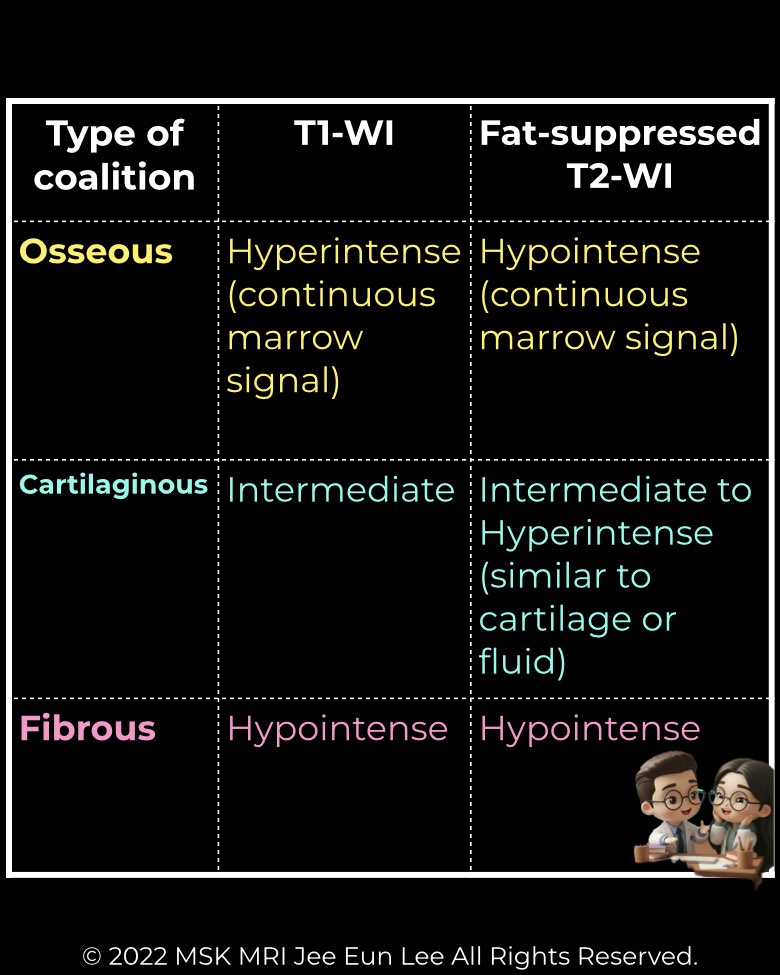
A fibrous coalition (syndesmosis) is a non-osseous union of tarsal bones bridged by dense fibrous tissue. MRI is superior to CT in detecting this subtle type.
MRI appearance
- Bridging tissue shows low signal intensity on all pulse sequences.
- Bones are abnormally close but separated by this thin, dark fibrous band.
- The articulation often looks narrowed or indistinct.
Anatomic features
- The interval between rudimentary facets may be widened but lacks normal cartilage.
- Frequently involves the middle subtalar joint, but can occur elsewhere.
- Differentiation from the medial talocalcaneal ligament is critical:
Radiology perspective
Fibrous coalitions are easily overlooked on plain radiographs but are well depicted on MRI due to their uniformly low signal bridge and associated bone changes. Their recognition is crucial in symptomatic adolescents and young adults with unexplained hindfoot pain.
#Radiology, #MSKMRI, #FibrousCoalition, #SubtalarJoint, #CoalitionImaging, #FootMRI, #OrthopedicImaging, #RadiologyEducation, #MSKImaging, #RadiologistLife
Visualizing MSK Radiology: A Practical Guide to Radiology Mastery
© 2022 MSK MRI Jee Eun Lee All Rights Reserved.
No unauthorized reproduction, redistribution, or use for AI training.