https://youtube.com/shorts/uzso2bRylAg
 Coalition by MSKMRI JEE EUN LEE.pdf
6.67MB
Coalition by MSKMRI JEE EUN LEE.pdf
6.67MB
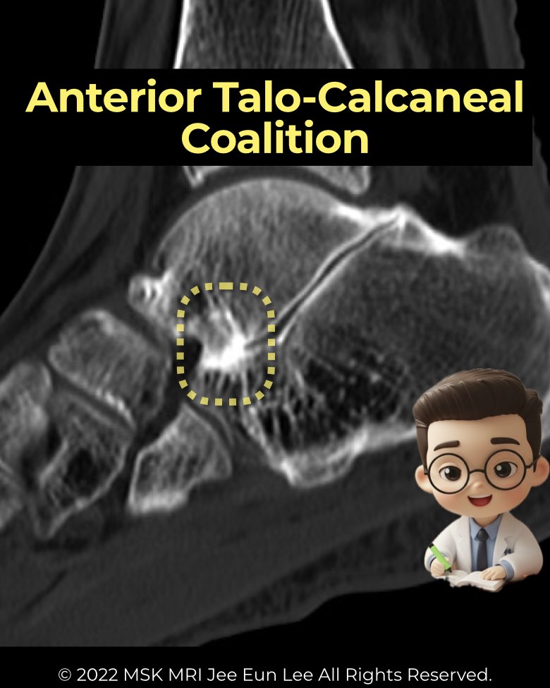
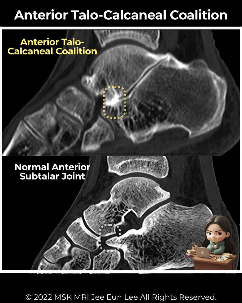
Coalition involving the anterior subtalar joint is exceptionally rare compared with middle or posterior types.
Anatomy & morphology
- The anterior facet is the smallest and most variable of the three subtalar facets.
- When a coalition occurs here, it almost always coexists with a middle facet coalition rather than appearing in isolation.
- Because of its small size and variable presence, anterior facet coalitions can be easily overlooked.
Imaging findings
- Radiographs: very limited role; no reliable indirect signs.
- CT & MRI:
Clinical relevance
- Isolated anterior coalitions are so uncommon that most literature describes them only as coexisting lesions with middle facet coalitions.
- Clinical presentation is indistinguishable from other talocalcaneal coalitions (hindfoot pain, stiffness, rigid flatfoot).
- Recognition is important, as involvement of the anterior facet may increase coalition extent and surgical complexity.
#Radiology, #MSKMRI, #SubtalarJoint, #AnteriorFacet, #CoalitionImaging, #FootMRI, #OrthopedicImaging, #RadiologyEducation, #MSKImaging, #RadiologistLife
Visualizing MSK Radiology: A Practical Guide to Radiology Mastery
© 2022 MSK MRI Jee Eun Lee All Rights Reserved.
No unauthorized reproduction, redistribution, or use for AI training.