https://youtube.com/shorts/cVAfMtvaLq0
 Coalition by MSKMRI JEE EUN LEE.pdf
6.67MB
Coalition by MSKMRI JEE EUN LEE.pdf
6.67MB
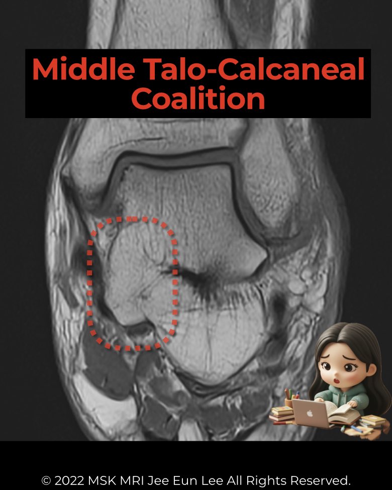
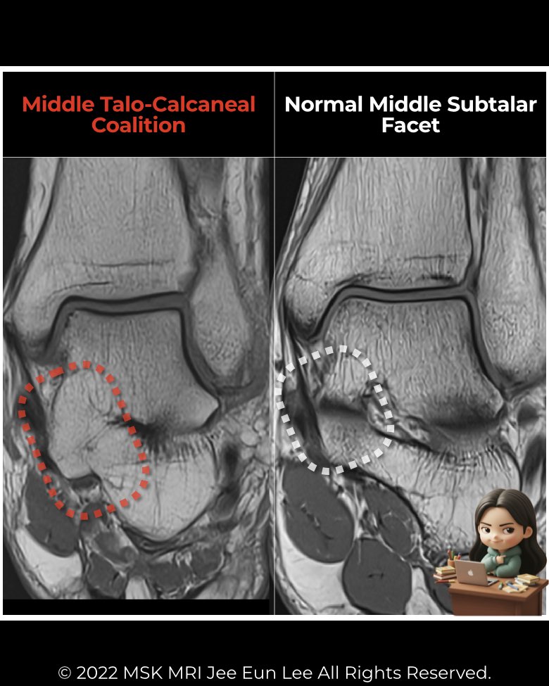
The middle subtalar joint is by far the most common site of talocalcaneal coalition.
Anatomy & morphology
- Occurs at the articulation between the sustentaculum tali of the calcaneus and the corresponding middle facet of the talus.
- Often involves the entire middle joint.
- Frequently associated with a dysmorphic sustentaculum tali (enlarged, rounded, or hypoplastic).
- On coronal MRI/CT: the normally horizontal or upward sloping facet instead slopes downward medially → the “drunken waiter sign.”
Imaging findings
- Radiographs (indirect signs):
- CT & MRI (definitive):
Clinical relevance
- Symptomatic presentation usually in adolescence (12–16 years).
- Restricted subtalar motion leads to pain, stiffness, rigid flatfoot, or peroneal spastic flatfoot.
- Recognition of this coalition type is essential since it represents the prototypical form of talocalcaneal coalition and has direct implications for surgical planning.
#Radiology, #MSKMRI, #SubtalarJoint, #MiddleFacet, #CoalitionImaging, #FootMRI, #OrthopedicImaging, #RadiologyEducation, #MSKImaging, #RadiologistLife
Visualizing MSK Radiology: A Practical Guide to Radiology Mastery
© 2022 MSK MRI Jee Eun Lee All Rights Reserved.
No unauthorized reproduction, redistribution, or use for AI training.