https://youtube.com/shorts/eAVK6PofZPk

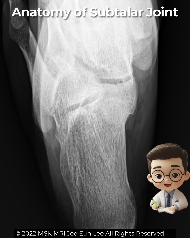
Harris Heel View: The Radiologist’s Shortcut to the Subtalar Joint
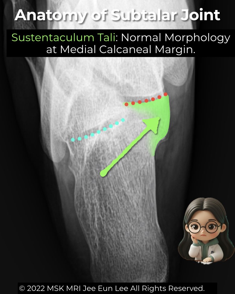
The Harris heel (axial) view is a specialized radiographic projection for the subtalar joint, especially the middle facet—key for detecting talocalcaneal coalition.
- Technique: patient standing with knee flexed or prone with dorsiflexed ankle; X-ray beam angled ~45° through the calcaneus.
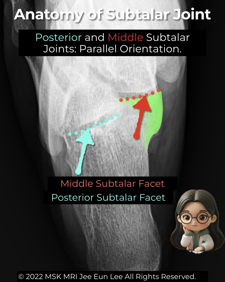
- Normal: sustentaculum tali is clear, posterior and middle facets appear parallel, anterior facet is obscured.
- Coalition clues: loss of facet parallelism, joint space narrowing, irregularity, osseous bar, or fibrocartilaginous cleft with sclerosis or cysts.
Though CT and MRI are now preferred for detailed evaluation, the Harris heel view still offers valuable insight when coalition is suspected but subtle on routine views.
#Radiology, #MSKMRI, #HarrisHeelView, #FootXray, #SubtalarJoint, #CoalitionImaging, #RadiologyEducation, #OrthopedicImaging, #MRIteaching, #RadiologistLife
Visualizing MSK Radiology: A Practical Guide to Radiology Mastery
© 2022 MSK MRI Jee Eun Lee All Rights Reserved.
No unauthorized reproduction, redistribution, or use for AI training.