https://youtube.com/shorts/bfZc7otMjQs

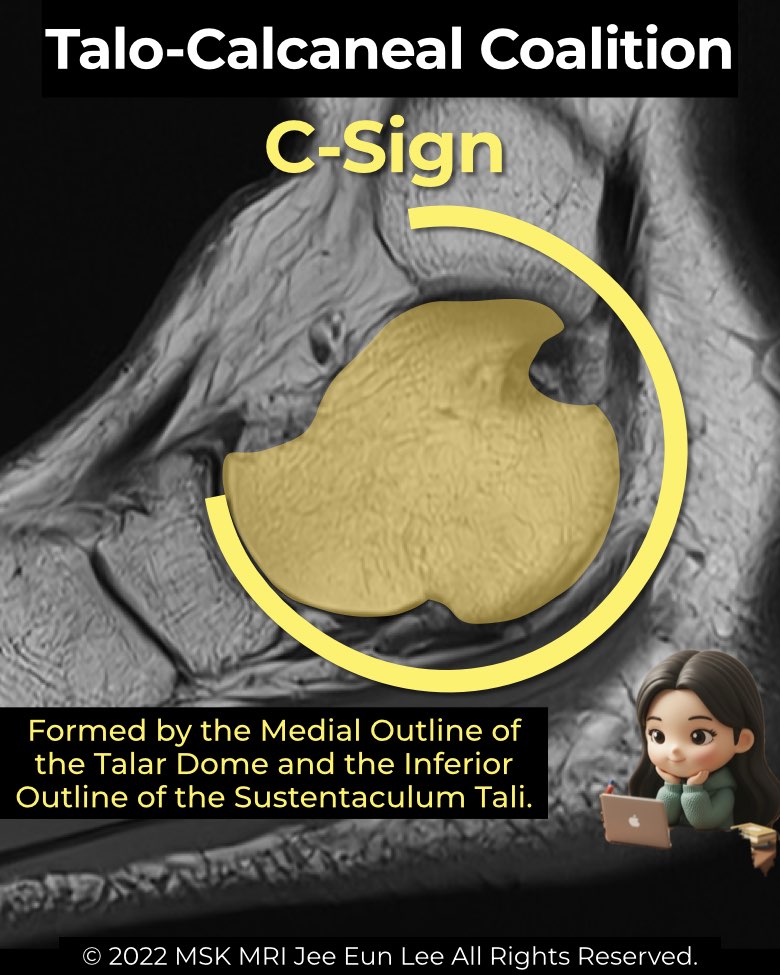
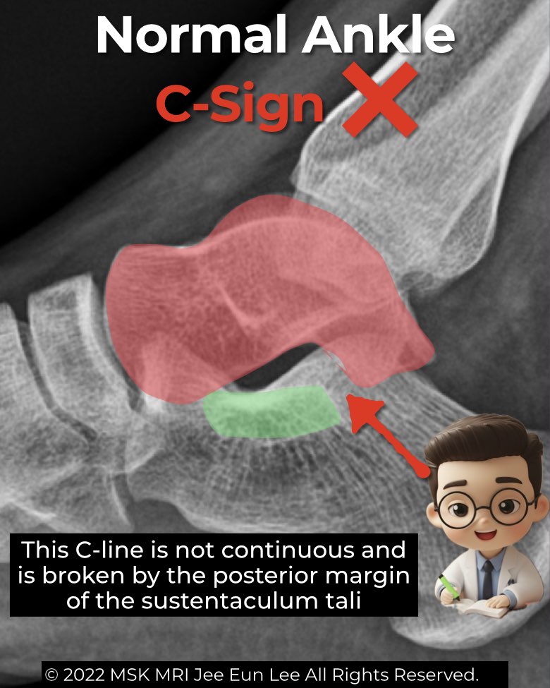
The C-sign is a well-known indirect radiographic marker of talocalcaneal coalition, best seen on weight-bearing lateral foot and ankle radiographs.
Anatomical basis
- Formed by a continuous C-shaped line connecting the medial talar dome with the inferior margin of the sustentaculum tali.
- In normal feet, this arc is broken at the posterior edge of the sustentaculum tali.
- Coalition of the middle or posterior facet creates the abnormal continuity.
Diagnostic utility
- Suggests talocalcaneal coalition, especially of the middle facet.
- Reported accuracy varies: sensitivity ranges from ~49–88%, specificity from ~87–91%.
- Often used with other secondary signs: talar beak, dysmorphic sustentaculum tali, or the absent middle facet sign.
Limitations & pitfalls
- Flatfoot (pes planus): can also produce a C-sign from close apposition of talus and sustentaculum tali, without coalition.
- Skeletal immaturity: sensitivity very low in children (<12 years, ~5%) compared with adults (~70%).
- Small coalitions: may not distort the outline enough to form the sign.
- Extra-articular posteromedial coalitions: typically do not show a C-sign, making them radiographically occult.
Radiology perspective
The C-sign remains a classic radiographic clue but must be interpreted in context. Correlation with CT or MRI is essential for confirmation, especially when evaluating young patients, flatfoot deformity, or suspected extra-articular coalitions.
#Radiology, #MSKMRI, #Csign, #TalocalcanealCoalition, #FootXray, #CoalitionImaging, #OrthopedicImaging, #RadiologyEducation, #MSKImaging, #RadiologistLife
Visualizing MSK Radiology: A Practical Guide to Radiology Mastery
© 2022 MSK MRI Jee Eun Lee All Rights Reserved.
No unauthorized reproduction, redistribution, or use for AI training.