https://youtube.com/shorts/212OVRW7qo8
 Coalition by MSKMRI JEE EUN LEE.pdf
6.67MB
Coalition by MSKMRI JEE EUN LEE.pdf
6.67MB
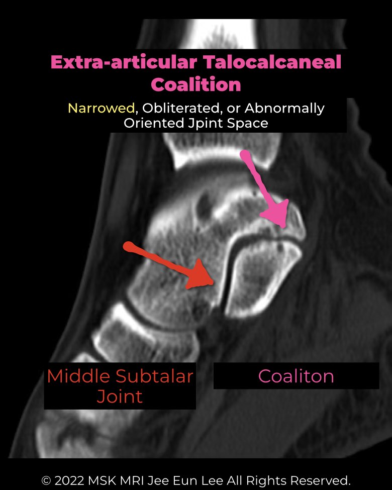
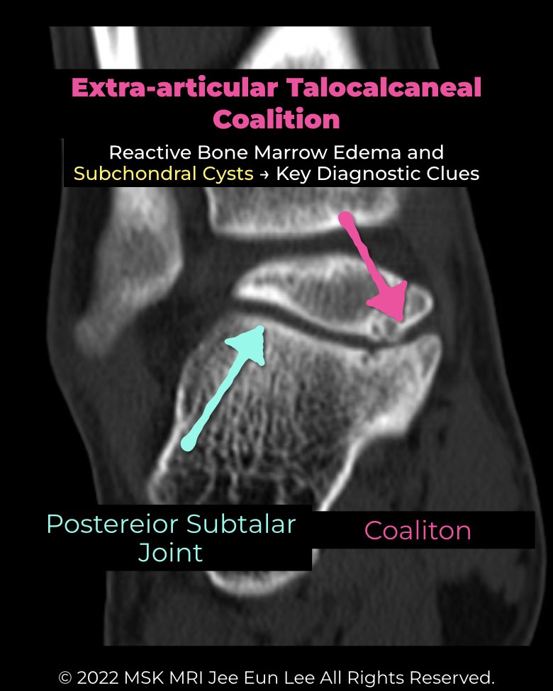
Definition & Location
- Abnormal union between talus and calcaneus outside the primary facets.
- Most common: posteromedial type (EA-PM) → between posterior sustentaculum tali and medial talar process.
- Rare: anterolateral variants.
Variants
- EA-PM coalition: ~28% of all TCC cases.
- With os sustentaculum: accessory ossicle integrates into coalition; reported in 16% of all coalitions / 24.1% of TCCs. Considered part of coalition, not just an accessory bone.
Imaging
- Radiographs: often occult; classic signs (C-sign, talar beak) usually absent. Clues: hypertrophy/prominence of posteromedial subtalar margin, visible os sustentaculum ossicle.
- CT: best for osseous detail; shows overgrowth, sclerosis, cysts, pseudoarthrosis. Defines os sustentaculum relationships.
- MRI: superior for non-osseous coalitions.
Clinical impact
- Tarsal Tunnel Syndrome (TTS): excrescences protrude into tunnel, compressing tibial nerve branches (esp. medial plantar nerve). MRI may show neuritis (edema, thickened nerve).
- Tendon pathology:
- Ganglion cysts: may arise from coalition and decompress into tunnel, worsening nerve compression.
Radiology perspective
- EA-TCC is often occult on radiographs, requiring CT/MRI for diagnosis.
- Key to differentiate from variants (accessory facet, medial talocalcaneal ligament): look for osseous deformity, irregular cortical margins, and marrow edema.
- MRI is essential to assess nerve compression, tendon pathology, and symptomatic mass effect.
#Radiology, #MSKMRI, #TalocalcanealCoalition, #ExtraArticularCoalition, #SubtalarJoint, #FootMRI, #TarsalTunnel, #RadiologyEducation, #OrthopedicImaging, #MSKImaging
Visualizing MSK Radiology: A Practical Guide to Radiology Mastery
© 2022 MSK MRI Jee Eun Lee All Rights Reserved.
No unauthorized reproduction, redistribution, or use for AI training.