https://youtube.com/shorts/abfILAu0hY0
 Coalition by MSKMRI JEE EUN LEE.pdf
6.67MB
Coalition by MSKMRI JEE EUN LEE.pdf
6.67MB
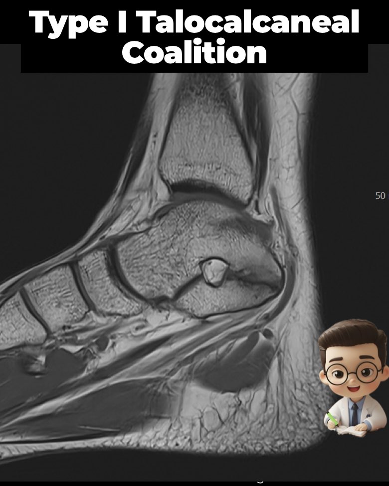
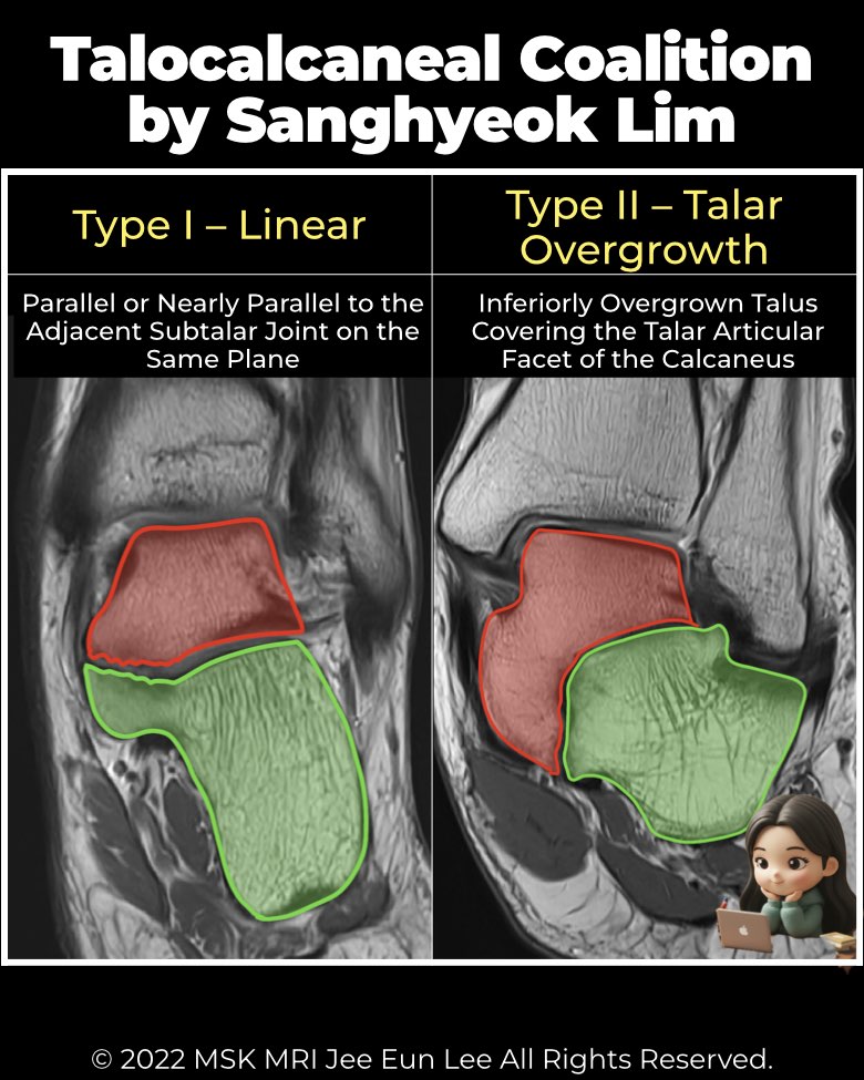
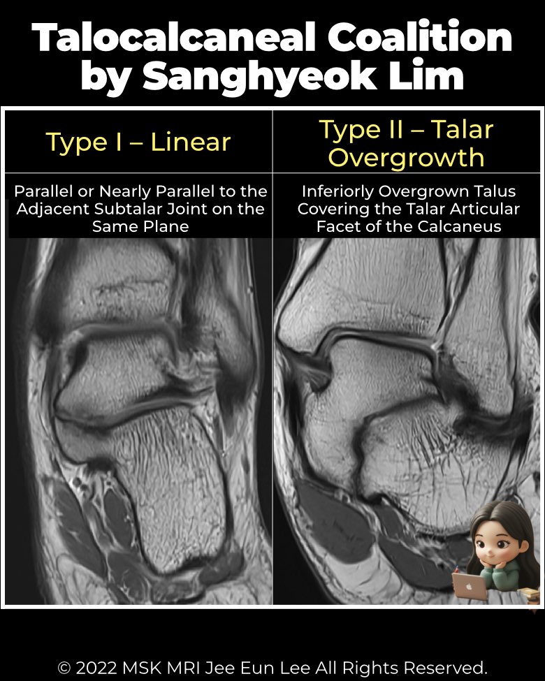
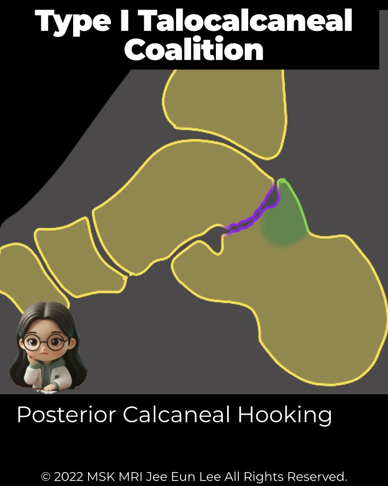
Definition & Morphology
- Linear coalition: parallel or nearly parallel to subtalar joint plane.
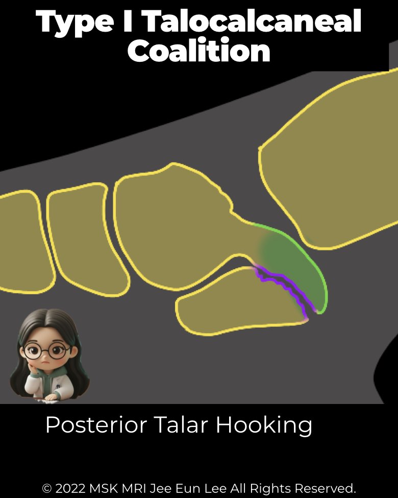
- May show posterior hooking:
Prevalence & Location
- Most common type – 64% (45/70 feet).
- Usually involves middle facet, but can extend to middle + posterior, or isolated posterior joint.
- All cases with posterior calcaneal hooking = posterior facet involvement.
Associated Findings
- Fracture fragments seen in 8 feet, especially with posterior hooking.
- More often from calcaneus (posterior calcaneal hooking) but can arise from talus.
- Important for surgeons: fragments may cause symptoms and often need removal.
Imaging role
- Best detected on coronal CT/MRI.
- Look for linear interface ± posterior hooking, fracture fragments, and facet involvement pattern.
#Radiology, #MSKMRI, #TalocalcanealCoalition, #SubtalarJoint, #CoalitionClassification, #FootMRI, #OrthopedicImaging, #RadiologyEducation, #MSKImaging, #RadiologistLife
Visualizing MSK Radiology: A Practical Guide to Radiology Mastery
© 2022 MSK MRI Jee Eun Lee All Rights Reserved.
No unauthorized reproduction, redistribution, or use for AI training.