https://youtube.com/shorts/-75F29oIHTE
 Coalition by MSKMRI JEE EUN LEE.pdf
6.67MB
Coalition by MSKMRI JEE EUN LEE.pdf
6.67MB
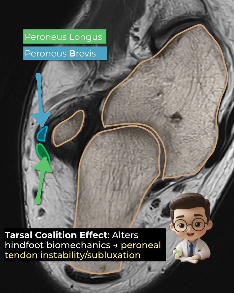
Mechanism
- Caused by hindfoot valgus malalignment from coalition, not direct impingement.
- Leads to posterior tibial tendon insufficiency → compensatory overactivity of peroneus brevis.
- Severe valgus → lateral hindfoot impingement → predisposes to peroneal tendon subluxation or dislocation.
Clinical clues
- Patients describe a popping sensation behind the lateral malleolus.
- Secondary findings: peroneal tenosynovitis, tendinosis, split tears, or complete ruptures.
Distinction from peroneal spasm
- Peroneal spastic flatfoot = peroneal muscles contract/shorten to stabilize a painful, stiff subtalar joint.
- It reflects protective spasm, not tendon instability.
- Tarsal coalition is a common cause, but other subtalar irritations can trigger it.
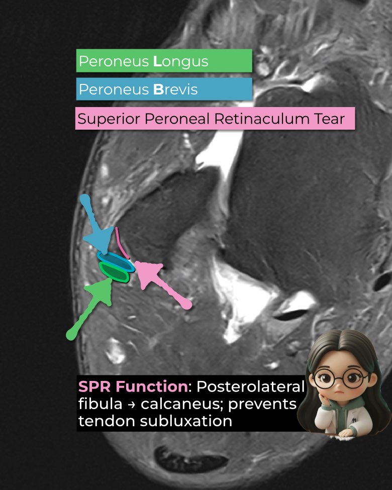
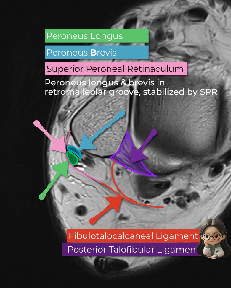
MRI Features
- Tendons partly out of fibular groove = subluxation; fully lateral/anterior = dislocation.
- SPR injury: detachment, pouching, periosteal elevation, avulsion (Oden I–IV).
- Coexisting longitudinal splits, groove dysplasia, retinacular thickening.
#Radiology, #MSKMRI, #PeronealTendon, #SPRinjury, #AnkleMRI, #CoalitionImaging, #RadiologyEducation, #OrthopedicImaging, #RadiologistLife
Visualizing MSK Radiology: A Practical Guide to Radiology Mastery
© 2022 MSK MRI Jee Eun Lee All Rights Reserved.
No unauthorized reproduction, redistribution, or use for AI training.