https://youtube.com/shorts/2GZJoxUmPZY
 Coalition by MSKMRI JEE EUN LEE.pdf
6.67MB
Coalition by MSKMRI JEE EUN LEE.pdf
6.67MB
Concept
- Developed for adult patients, using CT/MRI.
- Addresses morphologies (esp. calcaneal overgrowth) not covered by Rozansky’s pediatric system.
- Focus: coalition orientation, facet coverage, and osseous/cartilaginous nature.
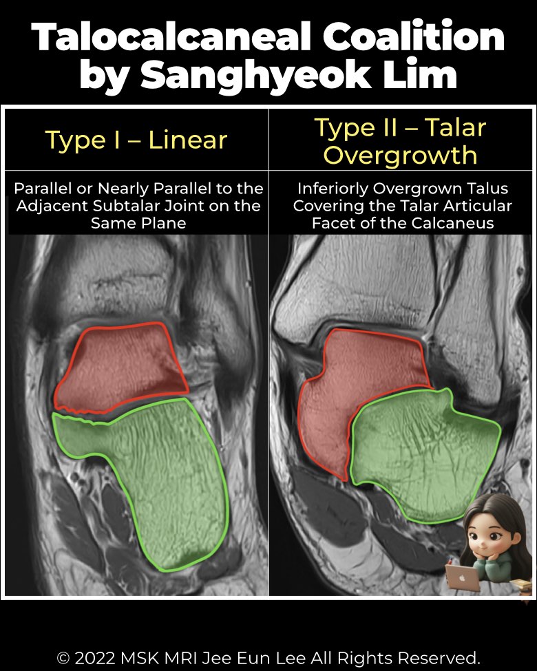
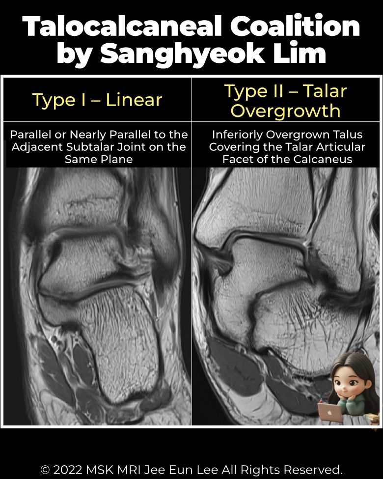
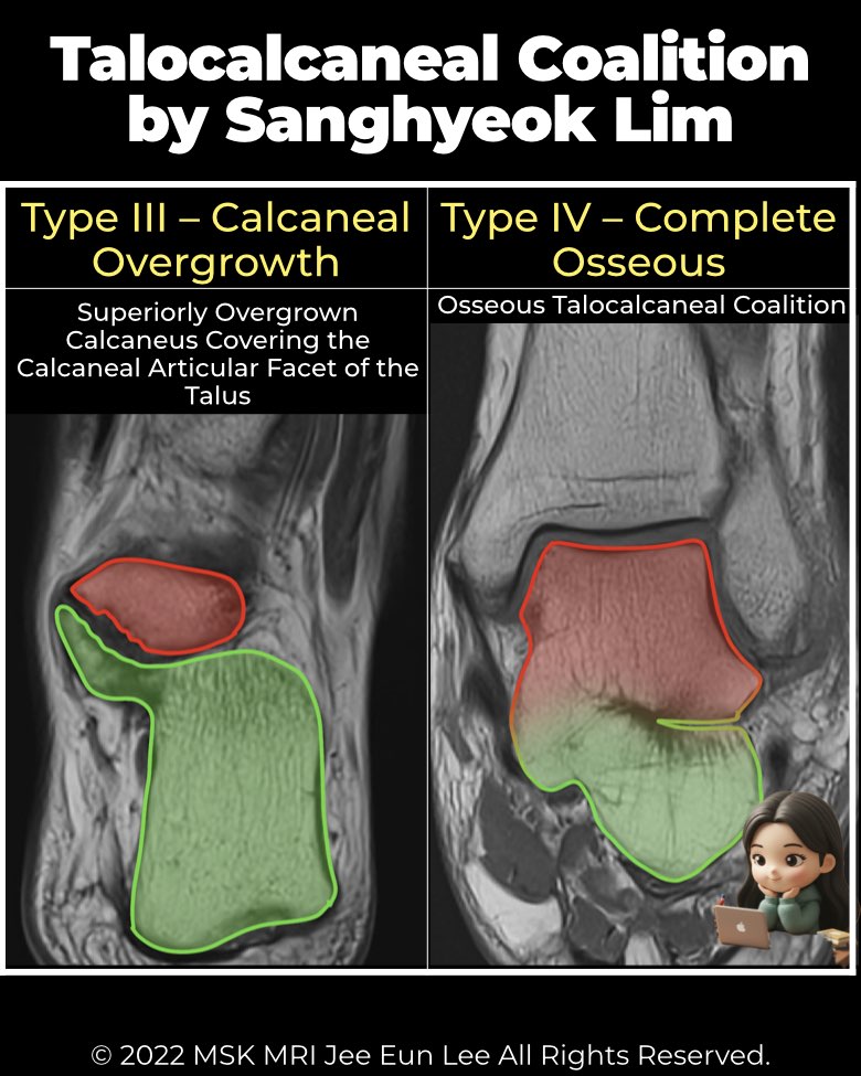
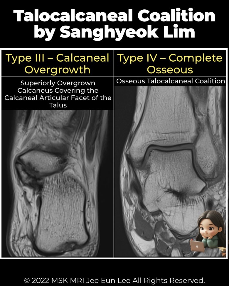
Types
- Type I: Linear – coalition parallel or nearly parallel to subtalar joint plane; may include posterior hooking. Most common (64%).
- Type II: Talar overgrowth – talus extends inferiorly over calcaneal facet (14%).
- Type III: Calcaneal overgrowth – calcaneus extends superiorly over talar facet (19%).
- Type IV: Complete osseous fusion – solid bony bar (3%).
Key associated findings
- Location: ~90% involve middle facet.
- Fracture fragments: seen in ~24%, especially Type I with calcaneal hooking and Type III. More often from calcaneus than talus.
- Clinical relevance: guides surgical planning (incision, approach, fragment removal).
Imaging role
- Coronal CT/MRI: best to define coalition type and detect subtle overgrowth or fracture fragments.
- Surgeons should be alert to calcaneal hooking and overgrowth patterns, which correlate with fragment risk.
#Radiology, #MSKMRI, #TalocalcanealCoalition, #SubtalarJoint, #FootMRI, #CoalitionClassification, #RadiologyEducation, #OrthopedicImaging, #MSKImaging, #RadiologistLife
Visualizing MSK Radiology: A Practical Guide to Radiology Mastery
© 2022 MSK MRI Jee Eun Lee All Rights Reserved.
No unauthorized reproduction, redistribution, or use for AI training.