https://youtube.com/shorts/gCEfG6gmCqU
“The 10-second rule every radiologist should know.”
✅ More structured MSK MRI guidance is available in my book,
Visualizing MSK Radiology: A Practical Guide to Radiology Mastery on Amazon.
https://www.amazon.com/dp/B0DJGMHMFS
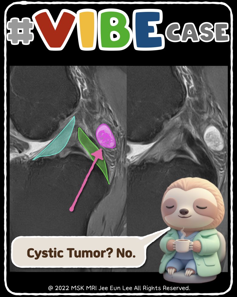
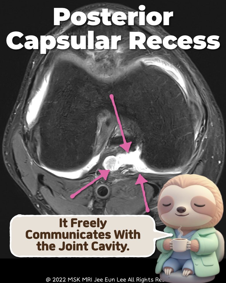
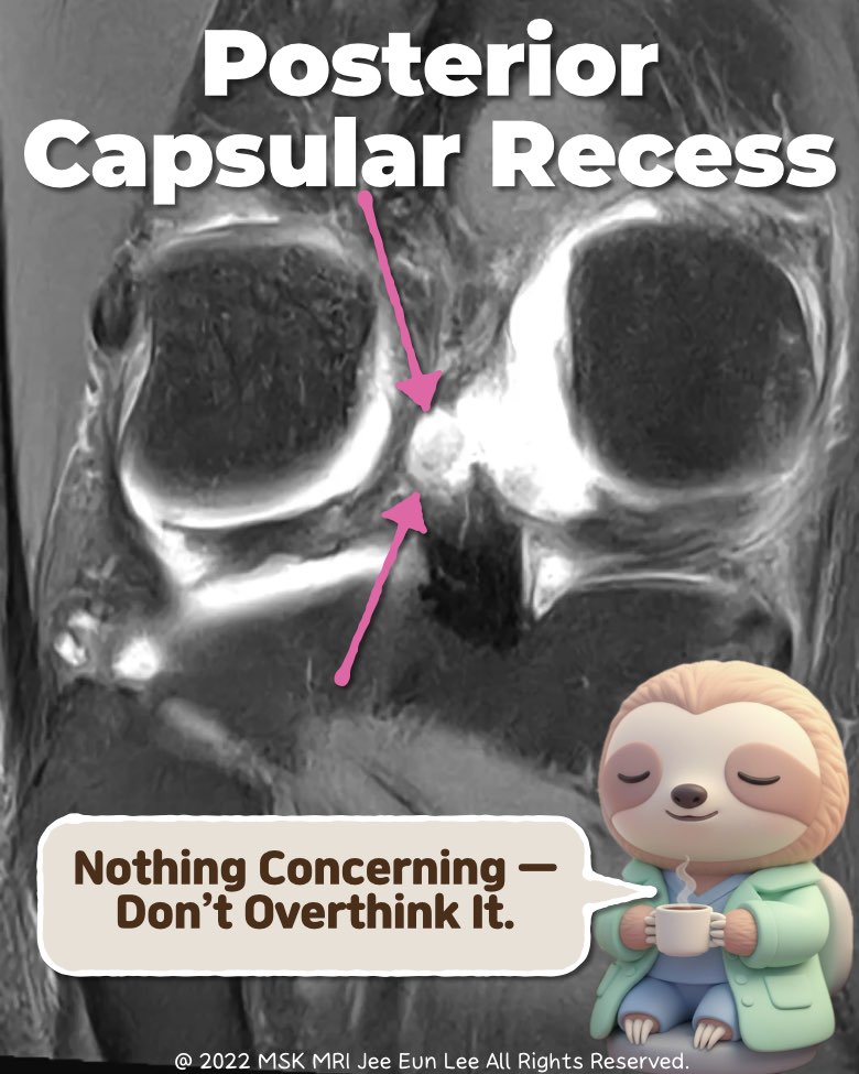
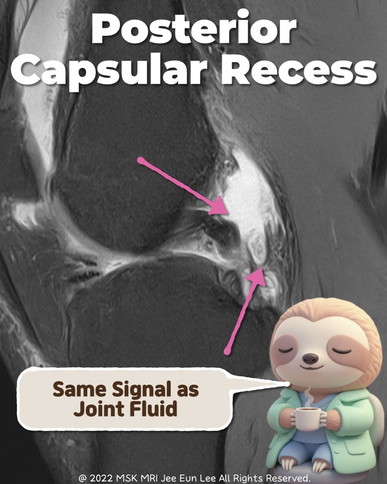
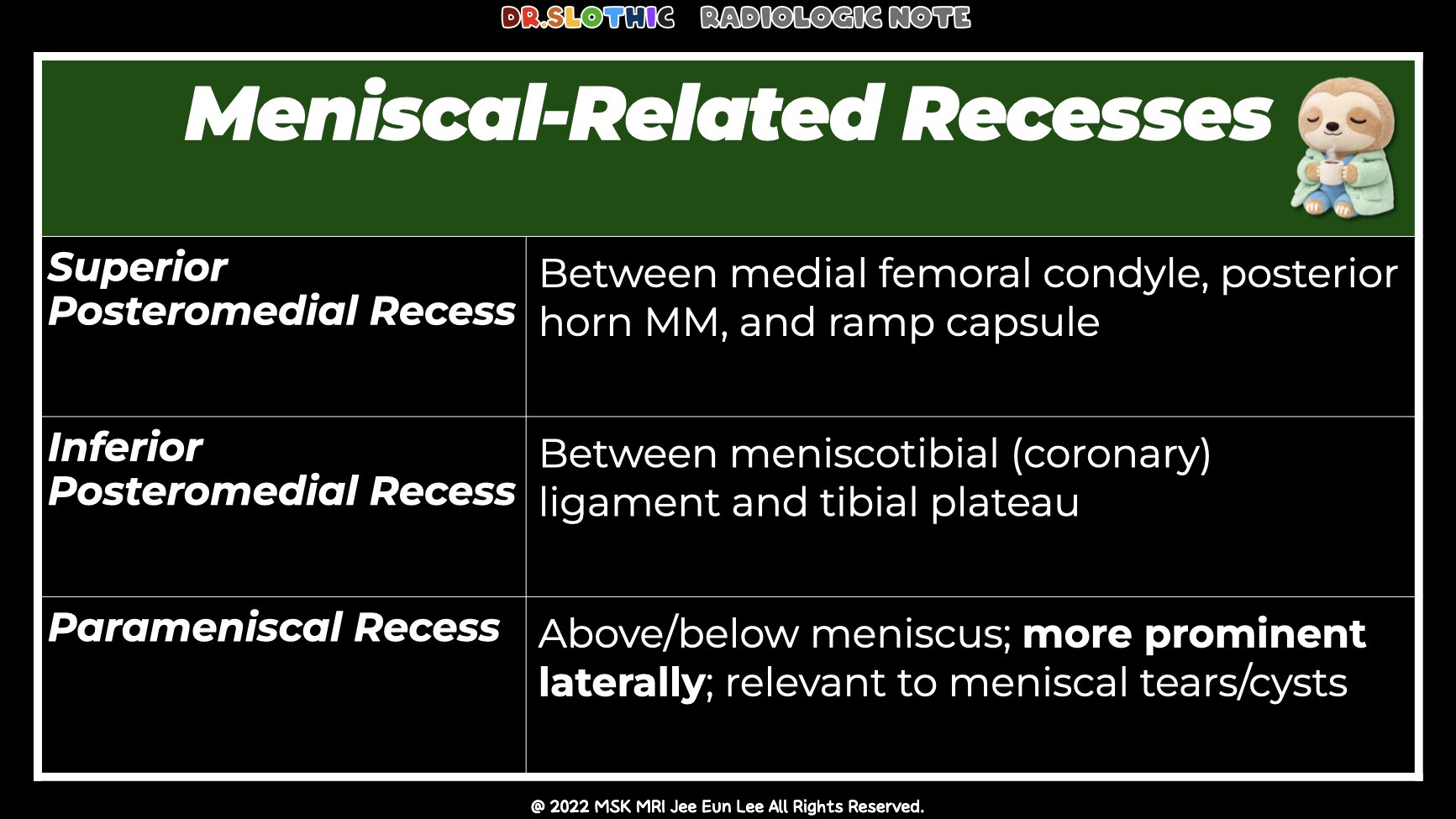
📌 Posterior Capsular Recess vs PCL Ganglion — Ultra Short Guide
1) Location (Start Here)
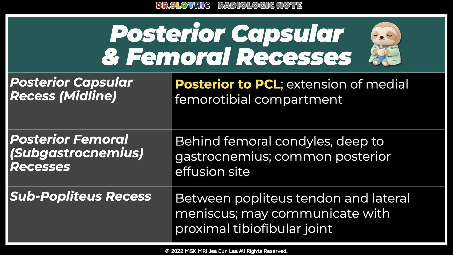
Recess: Posteromedial, behind the joint capsule, deep to the medial gastrocnemius
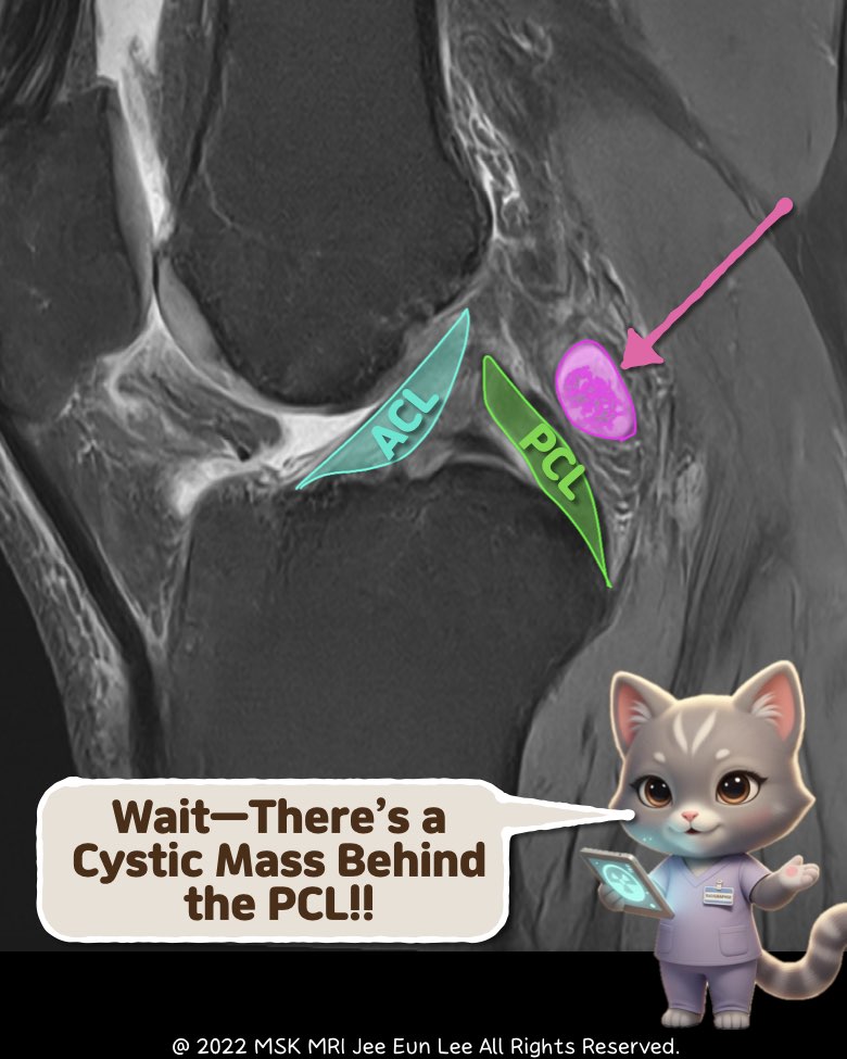
PCL Cyst: Intercondylar notch, around the PCL or between cruciate fibers
2) Morphology
Recess: Flat crescent-shaped fluid, widely communicating with the joint
PCL Cyst: Rounded or multilobulated, well-defined discrete fluid mass
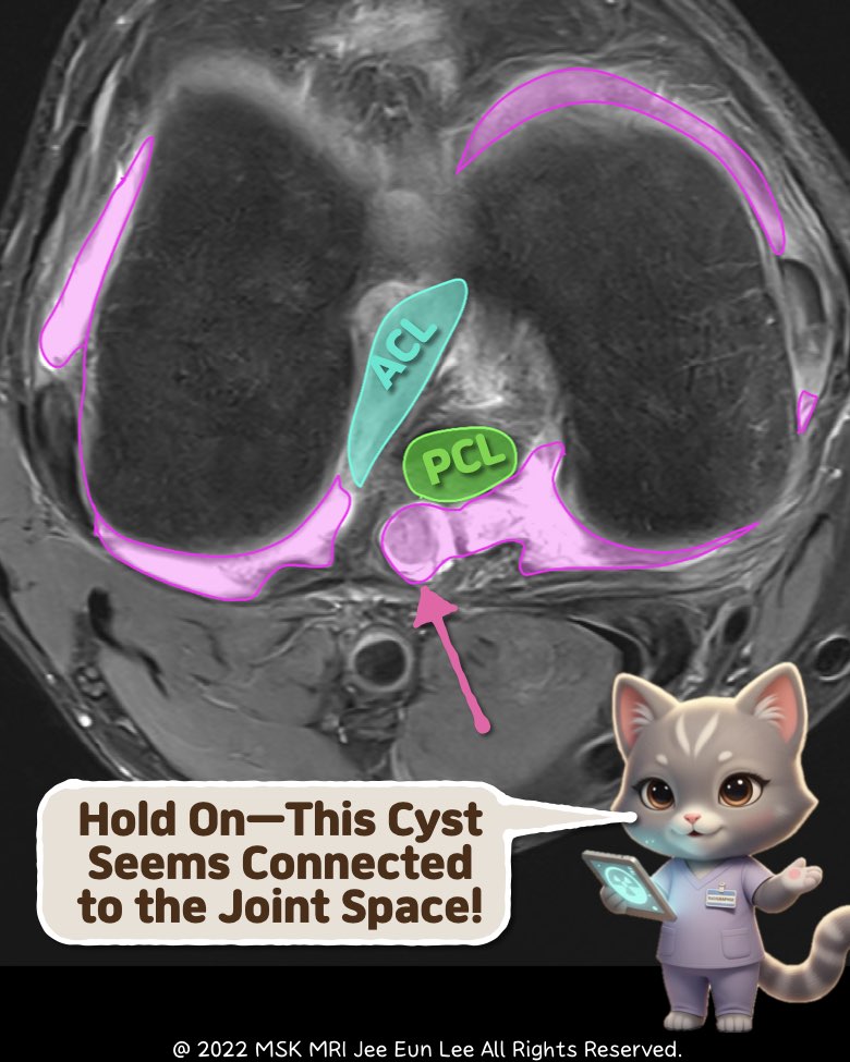
3) Joint Communication (Most Important)
Recess: Clearly communicates with the joint space
PCL Cyst: No true communication; size independent of effusion
4) Signal & Structure
Recess: Simple fluid signal, thin wall, no septations
PCL Cyst: T2 bright, internal septations common, possible wall thickening
5) One-Line Differentiation
Recess → Posteromedial + joint-connected fluid
PCL Cyst → Intercondylar + PCL-attached mass
#MSKRadiology, #KneeMRI, #PCL, #GanglionCyst, #RadiologyTips, #MRIRecess, #Orthopedics, #RadiologyEducation, #DrSlothic, #ImagingPearls
Visualizing MSK Radiology: A Practical Guide to Radiology Mastery
© 2022 MSK MRI Jee Eun Lee All Rights Reserved.
No unauthorized reproduction, redistribution, or use for AI training.
'✅ Dr. Slothic Notes' 카테고리의 다른 글
| 📌 When the MPFL Tears Without Patellar Dislocation (0) | 2025.11.23 |
|---|---|
| 📌 MPFL: It’s Not One Ligament. (0) | 2025.11.23 |
| 📌 Key Imaging Features Suggesting Neurogenic Origin (PNSTs) (0) | 2025.11.23 |
| 📌 The Easiest Way to Avoid Misdiagnosing PCL Tears (0) | 2025.11.23 |
| 📌 Hemangioma vs Fracture: The 5-Second MRI Trick. (0) | 2025.11.22 |