✅ More structured MSK MRI guidance is available in my book,
Visualizing MSK Radiology: A Practical Guide to Radiology Mastery on Amazon.
https://www.amazon.com/dp/B0DJGMHMFS
https://youtube.com/shorts/cK9u_xGonrk
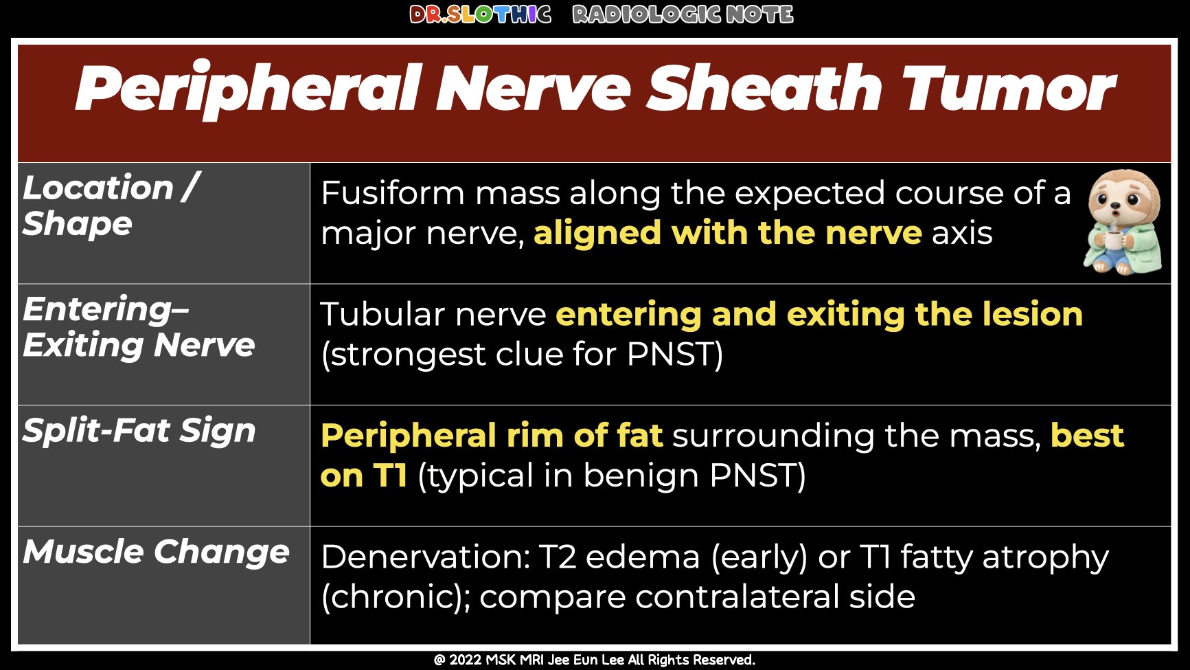
1) Location Along Major Nerves
Masses located along the typical course of major nerves
(e.g., median nerve, sciatic nerve, tibial nerve)
should immediately raise suspicion for a neurogenic tumor.
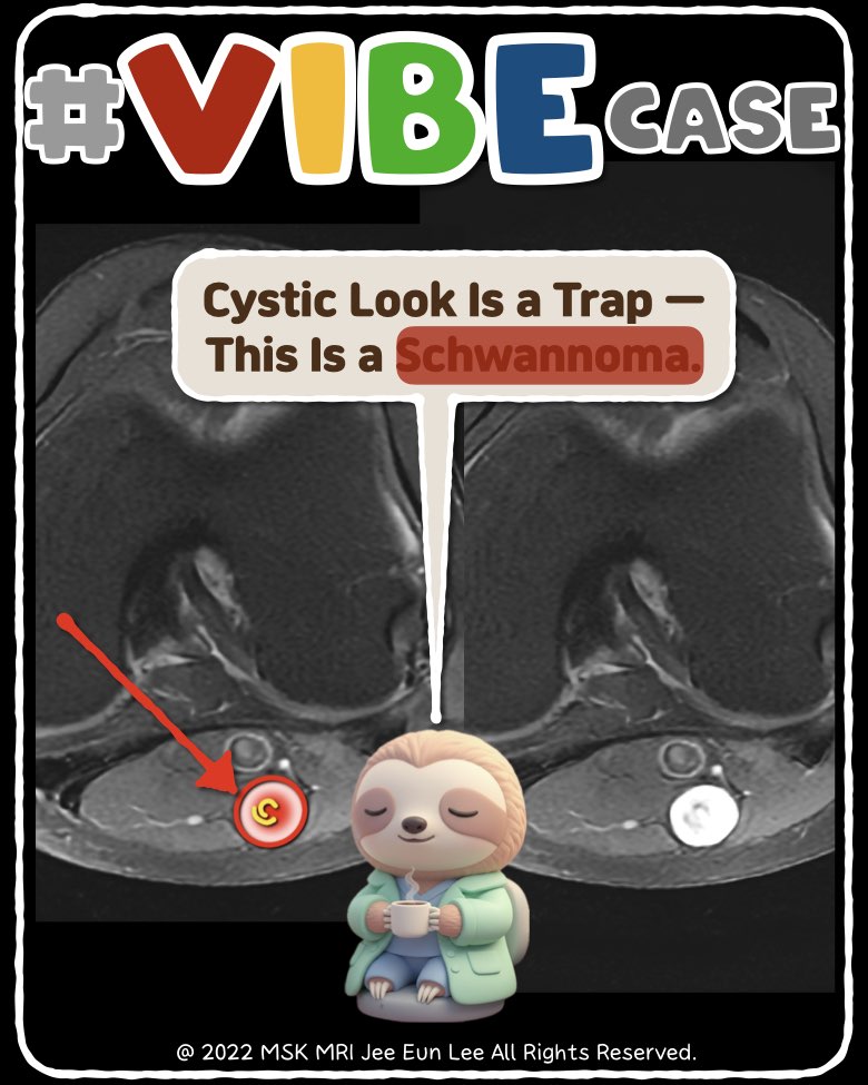
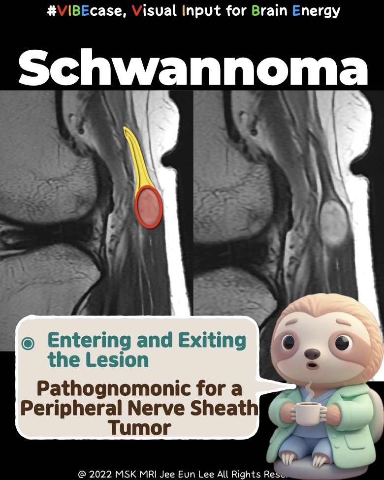
2) Entering and Exiting Nerve
One of the strongest clues:
Visualization of a tubular nerve entering and exiting the mass.
This finding is considered pathognomonic for Peripheral Nerve Sheath Tumors (PNSTs).
3) Fusiform Shape
Lesions that are fusiform (spindle-shaped)
—elongated along the nerve’s axis—
are characteristic of neurogenic neoplasms and rare in soft tissue sarcomas.
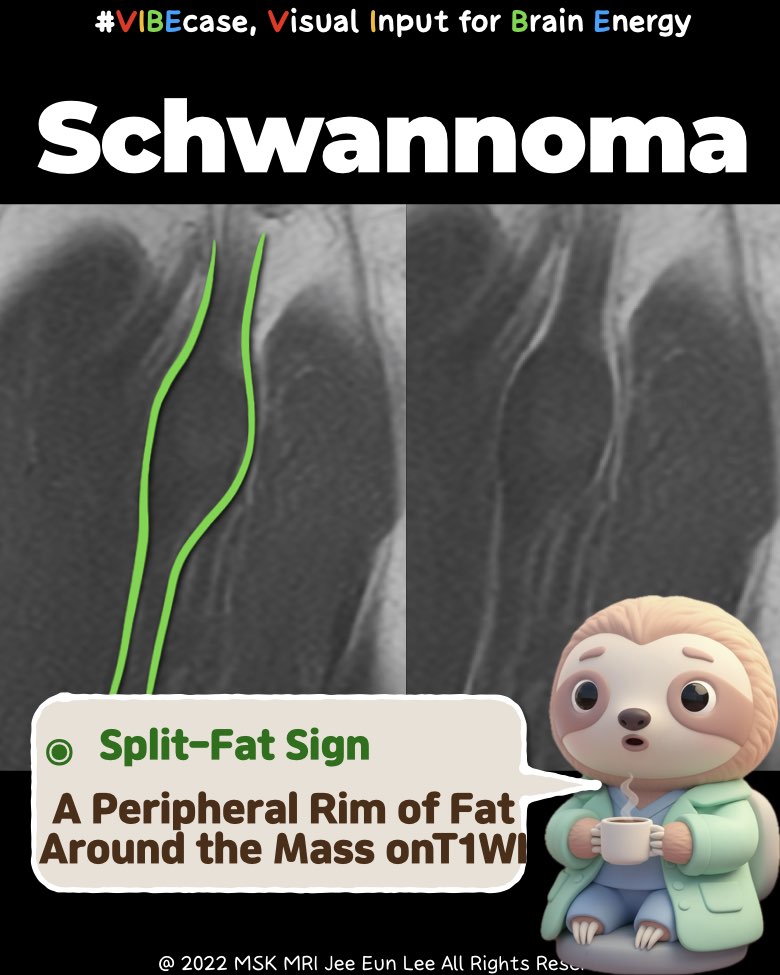
4) Split-Fat Sign
A rim of fat surrounding the mass
→ known as the split-fat sign.
This suggests the tumor originated in the intermuscular fat plane
near the neurovascular bundle.
Best visualized on T1-weighted MRI.
Common in benign PNSTs.
5) Muscle Abnormalities
Changes in muscles supplied by the affected nerve can reinforce the diagnosis:
- Fatty atrophy or decreased muscle bulk → best seen on T1
- Edematous muscle changes from early denervation → best seen on T2
- Always compare with the contralateral side for subtle cases.
- Visualizing MSK Radiology: A Practical Guide to Radiology Mastery
© 2022 MSK MRI Jee Eun Lee. All Rights Reserved.
No unauthorized reproduction, redistribution, or use for AI training.
#MedicalImaging, #Radiology, #MRI, #PNST, #NerveSheathTumor, #Schwannoma, #NeurogenicTumor, #TibialNerve, #MSKRadiology, #DrSlothic
'✅ Dr. Slothic Notes' 카테고리의 다른 글
| 📌 When the MPFL Tears Without Patellar Dislocation (0) | 2025.11.23 |
|---|---|
| 📌 MPFL: It’s Not One Ligament. (0) | 2025.11.23 |
| 📌 Posterior Capsular Recess vs PCL Ganglion — Ultra Short Guide (0) | 2025.11.23 |
| 📌 The Easiest Way to Avoid Misdiagnosing PCL Tears (0) | 2025.11.23 |
| 📌 Hemangioma vs Fracture: The 5-Second MRI Trick. (0) | 2025.11.22 |