https://youtube.com/shorts/C9JzEoyZF1o
✅ More structured MSK MRI guidance is available in my book,
Visualizing MSK Radiology: A Practical Guide to Radiology Mastery on Amazon.
https://www.amazon.com/dp/B0DJGMHMFS
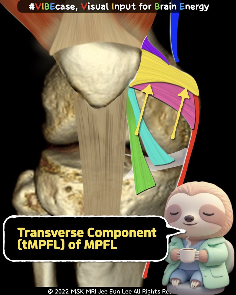
1) MPFL = TWO Components
Not one band, but a two-part complex:
- Transverse component
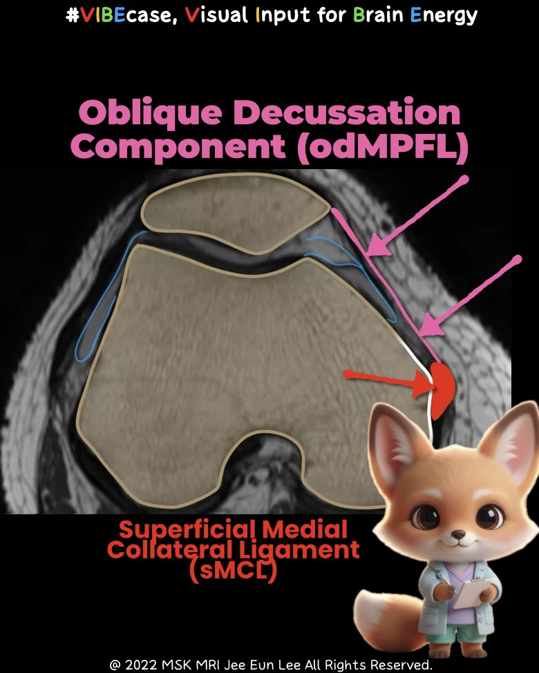
• From the medial femoral epicondyle → adductor tubercle - Oblique decussation component
• From the superficial MCL
2) Common Insertion
Both components merge and insert onto the medial patella
(plus deep fibers of the vastus medialis).
3) Key MRI Tips
- The transverse portion is thin → often subtle or hard to see
- The oblique portion frequently shows edema or tearing
→ even without lateral patellar dislocation - Superficial MCL injury?
→ Check the oblique MPFL component (high association)
🎯 Dr. Slothic’s One-Line Pearl
“The MPFL is a two-part complex, and the oblique portion tears more often than you expect— even without patellar dislocation.”
#MSKRadiology, #KneeMRI, #MPFL, #PatellarInstability, #RadiologyEducation, #Orthopedics, #Patellofemoral, #MRIAnatomy, #DrSlothic, #ImagingPearls
Visualizing MSK Radiology: A Practical Guide to Radiology Mastery
© 2022 MSK MRI Jee Eun Lee All Rights Reserved.
No unauthorized reproduction, redistribution, or use for AI training.
#MPFL, #KneeLigament, #sMCL, #KneeMRI, #FemoralAttachment, #KneeAnatomy, #Orthopedics, #LigamentInjury, #MedicalImaging


'✅ Dr. Slothic Notes' 카테고리의 다른 글
| 📌 RTSA Glenoid Complications: The Essentials (0) | 2025.12.01 |
|---|---|
| 📌 When the MPFL Tears Without Patellar Dislocation (0) | 2025.11.23 |
| 📌 Key Imaging Features Suggesting Neurogenic Origin (PNSTs) (0) | 2025.11.23 |
| 📌 Posterior Capsular Recess vs PCL Ganglion — Ultra Short Guide (0) | 2025.11.23 |
| 📌 The Easiest Way to Avoid Misdiagnosing PCL Tears (0) | 2025.11.23 |