https://youtube.com/shorts/FEbTVsKoT1Y
How to Differentiate Normal Remodeling from True Mechanical Failure
Radiographic and CT follow-up are the backbone of evaluating glenoid-sided complications after Reverse Total Shoulder Arthroplasty (RTSA).
A few key imaging patterns reliably separate benign postoperative changes from worrisome loosening or migration.
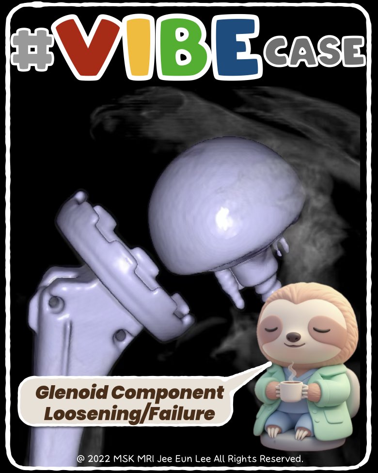
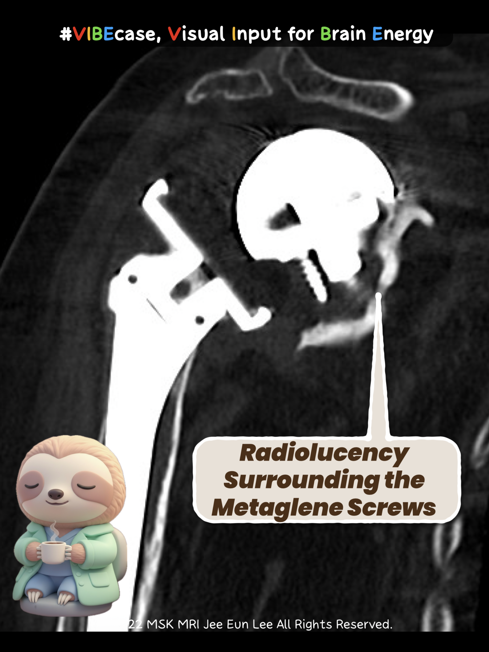
1. Glenoid Loosening – The Red Flags
What to look for:
- Progressive radiolucent lines >2 mm at the baseplate–bone or screw–bone interface
- Circumferential lucency or interval increase on follow-up
- Screw haloing or breakage
- Positional change of the baseplate (tilt, subsidence)
- Periprosthetic osteolysis
Best modalities:
Serial radiographs → CT for lucent rims & bone loss → SPECT/CT for painful prostheses
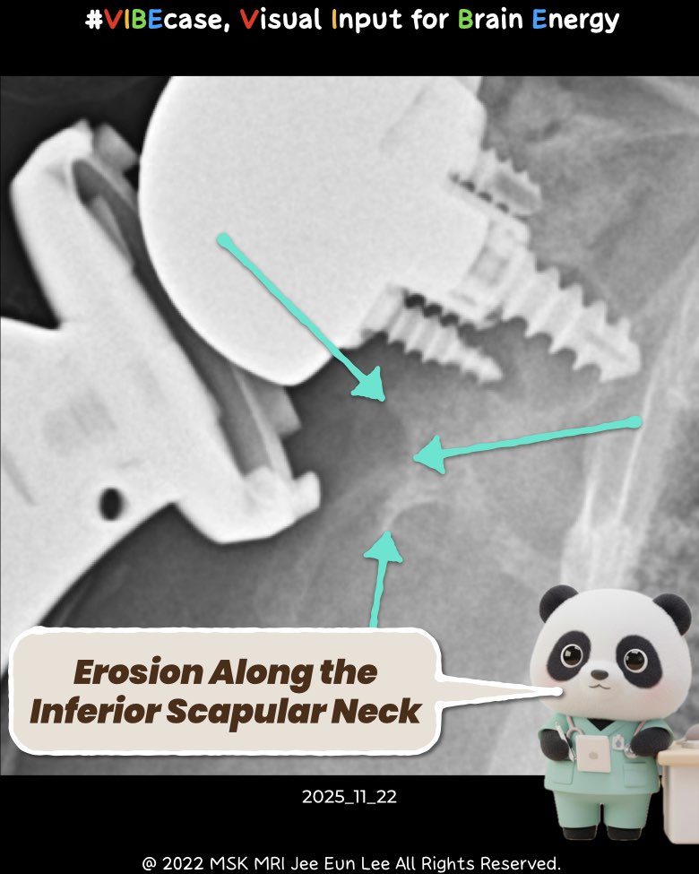
2. Scapular Notching – Expected vs. Problematic
Definition:
Inferomedial cortical erosion at the inferior scapular neck due to adduction impingement.
Imaging appearance:
- Concave defect just below the baseplate (Sirveaux grades 1–4)
- CT: sharp cortical step-off, cavitary loss ± osteolysis from polyethylene wear
- SPECT/CT may show focal uptake when symptomatic
3. Glenoid Migration & Tilt – When Position Starts to Drift
What suggests mechanical failure:
- Cranial or caudal translation of baseplate/glenosphere
- Medialization or lateralization compared with initial postoperative imaging
- Development of unintended superior tilt
- Progressive change beyond expected early settling
Best modalities:
Serial standardized radiographs → CT for precise translation/rotation assessment
Quick Summary for Clinicians
| Complication | Key Imaging Signs | Best Modality |
| Glenoid loosening | >2 mm radiolucent lines, screw haloing, osteolysis, positional change | Radiographs, CT, SPECT/CT |
| Scapular notching | Inferomedial cortical erosion, graded severity, sub-baseplate bone loss | AP radiographs, CT |
| Migration/tilting | Cranial/caudal shift, unintended superior tilt, progressive rotation | Radiographs, CT |
#RTSA, #ShoulderArthroplasty, #GlenoidLoosening, #ScapularNotching, #Orthopaedics, #Radiology, #MSKRadiology, #ShoulderImaging, #CTImaging, #PostoperativeImaging
Visualizing MSK Radiology: A Practical Guide to Radiology Mastery
© 2022 MSK MRI Jee Eun Lee All Rights Reserved.
No unauthorized reproduction, redistribution, or use for AI training.


'✅ Dr. Slothic Notes' 카테고리의 다른 글
| 📌 CT-Negative, MRI-Positive: The Cancellous Stress Fracture Pattern You Should Not Miss (0) | 2025.12.03 |
|---|---|
| 📌 Coracoid Stress Fracture After Reverse Total Shoulder Arthroplasty (0) | 2025.12.02 |
| 📌 When the MPFL Tears Without Patellar Dislocation (0) | 2025.11.23 |
| 📌 MPFL: It’s Not One Ligament. (0) | 2025.11.23 |
| 📌 Key Imaging Features Suggesting Neurogenic Origin (PNSTs) (0) | 2025.11.23 |