https://youtube.com/shorts/WfY4j4MNEHQ
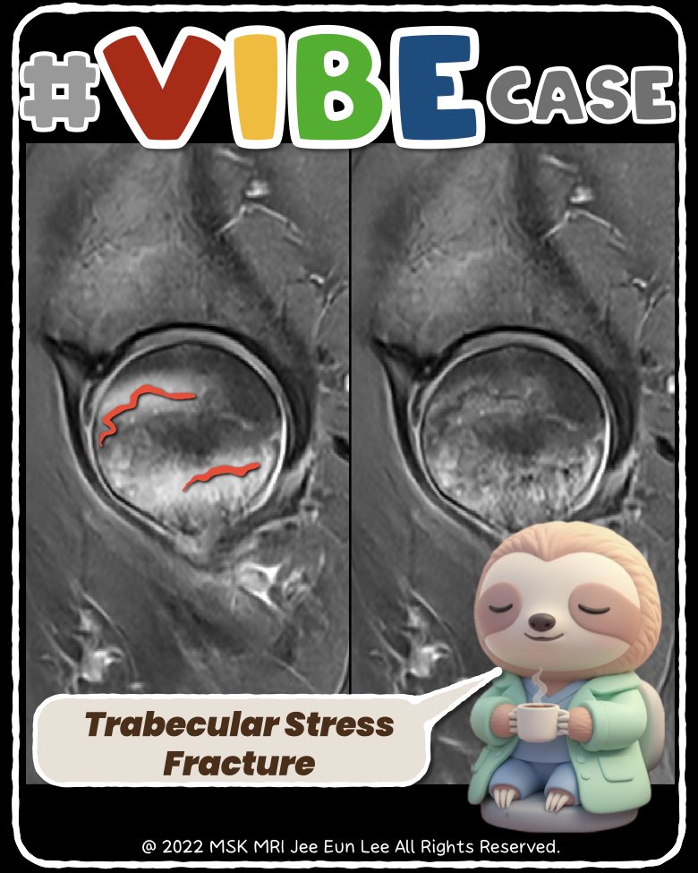
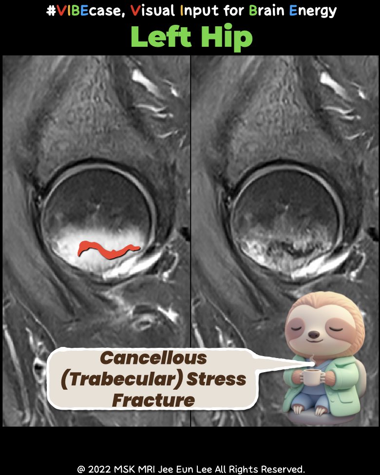
When the Cortex Is Intact but the Trabecula Collapses: Why Only MRI Reveals the True Injury
Cancellous (trabecular) stress injuries of the femoral neck and femoral head often remain completely invisible on CT.
The cortex stays intact, the subchondral contour appears normal, and the subtle attenuation changes fall below CT’s detection threshold.
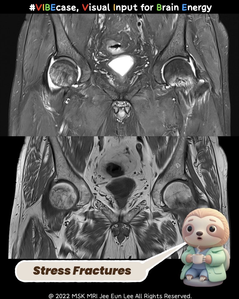
MRI, however, shows the full pathophysiology:
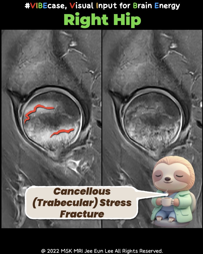
1. T1-weighted MRI
- Irregular, wavy, crumpled low-signal trabecular lines
- No cortical break, no subchondral crescent line
2. T2/STIR / PD-FS
- Prominent diffuse marrow edema
- Serpiginous dark microfracture lines within the edema
- No double-line sign and no collapse (distinguishes from AVN)
3. Mechanism
A single compression-dominant stress overload affecting the inferomedial femoral neck and the weight-bearing dome of the femoral head, producing a continuous-spectrum trabecular injury.
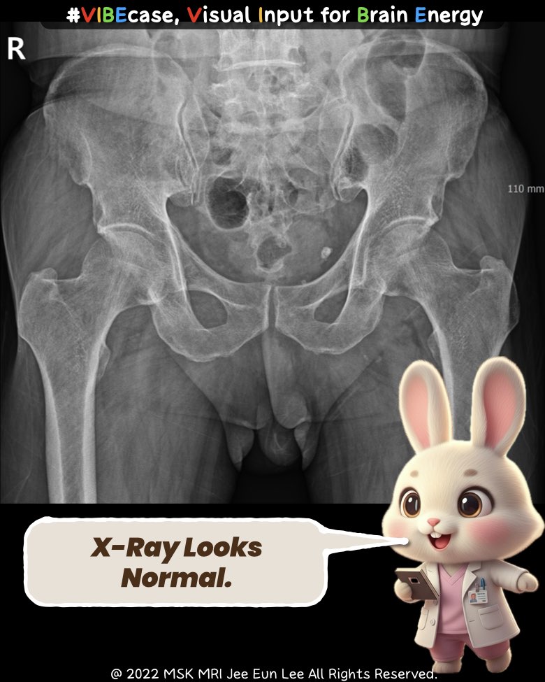
4. Why CT Fails
- Cortex intact → no structural cue
- Microtrabecular collapse below CT spatial resolution
- Marrow edema undetectable on CT
- Subtle density changes obscured by noise and beam hardening
- Articular surface remains morphologically normal
Conclusion
CT stays “normal,” but MRI reveals the biologic truth.
This pattern is a classic CT-occult, MRI-diagnostic cancellous stress fracture.
#StressFracture, #FemoralNeck, #FemoralHead, #MRIImaging, #MSKRadiology, #BoneMarrowEdema, #OccultFracture, #Orthopaedics, #RadiologyEducation, #CTvsMRI, #Vibecase,
Visualizing MSK Radiology: A Practical Guide to Radiology Mastery
© 2022 MSK MRI Jee Eun Lee All Rights Reserved.
No unauthorized reproduction, redistribution, or use for AI training.

'✅ Dr. Slothic Notes' 카테고리의 다른 글
| 📌 Understanding the Difference: Fifth Metatarsal Apophysis vs. Fracture (0) | 2025.12.06 |
|---|---|
| 📌 Insufficiency Fractures of the Unresurfaced Patella After TKA (0) | 2025.12.06 |
| 📌 Coracoid Stress Fracture After Reverse Total Shoulder Arthroplasty (0) | 2025.12.02 |
| 📌 RTSA Glenoid Complications: The Essentials (0) | 2025.12.01 |
| 📌 When the MPFL Tears Without Patellar Dislocation (0) | 2025.11.23 |