https://youtube.com/shorts/3PTgaZCGquo
https://open.spotify.com/episode/7wJqd534Vmqx8y6KBSkxwB?si=4hz_MM9ESk2H2WMWN593lQ
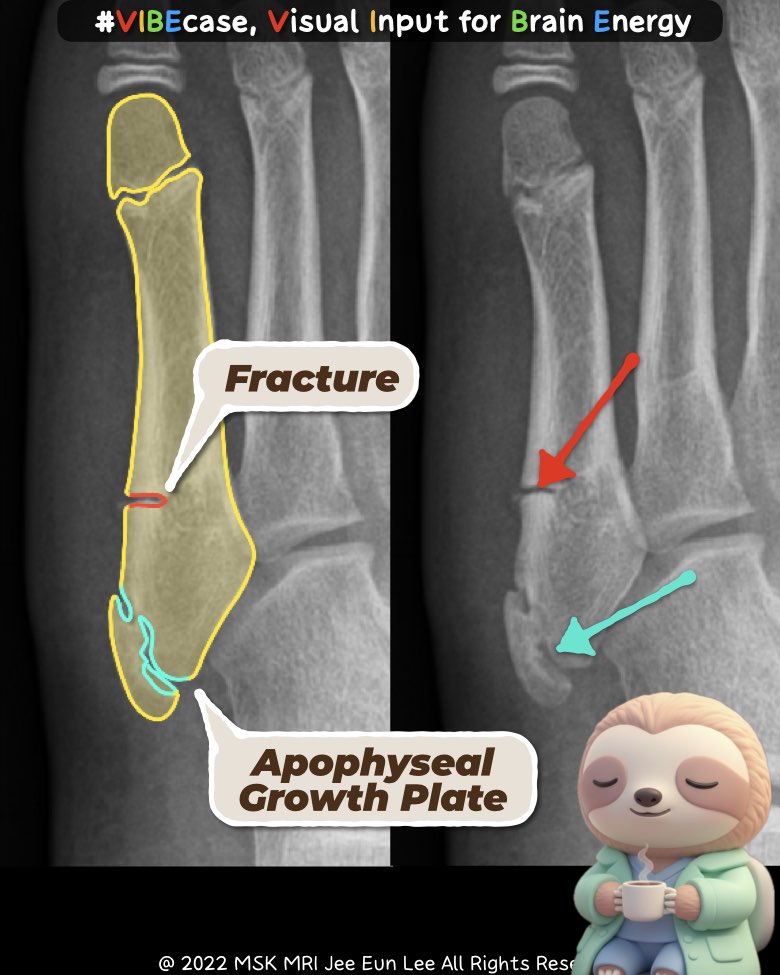
[Lecture] 📌 Understanding the Difference: Fifth Metatarsal Apophysis vs. Fracture
Dr. Slothic MSK Radiology Podcast · Episode
open.spotify.com
1. Why This Distinction Matters
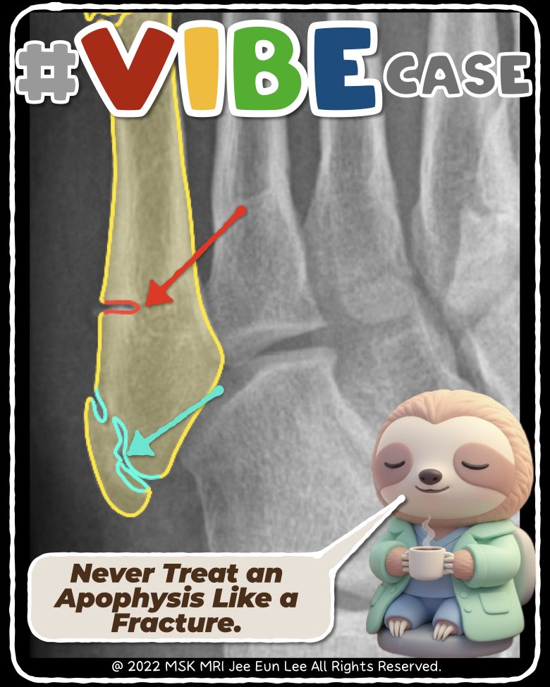
Fifth metatarsal apophysis is a normal secondary ossification center seen in children and adolescents, whereas a fracture represents a pathological break that follows trauma and often requires treatment.
Key differentiators include age, orientation on imaging, clinical context, and soft-tissue findings.
2. Age and Clinical Context
Apophysis
- Occurs in skeletally immature patients (girls ~9–11, boys ~11–14).
- Normal developmental variant with no definite trauma.
- May present as overuse pain (Iselin disease).
Fracture
- Occurs at any age, especially athletes.
- Typically follows acute inversion injury with sudden pain, swelling, or ecchymosis.
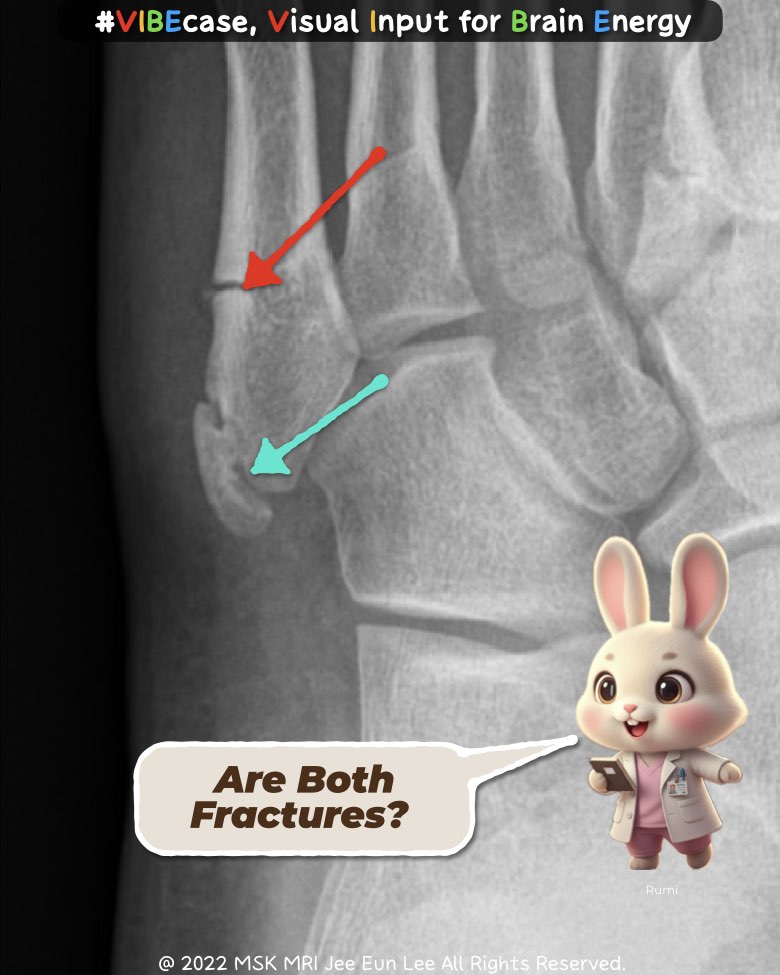
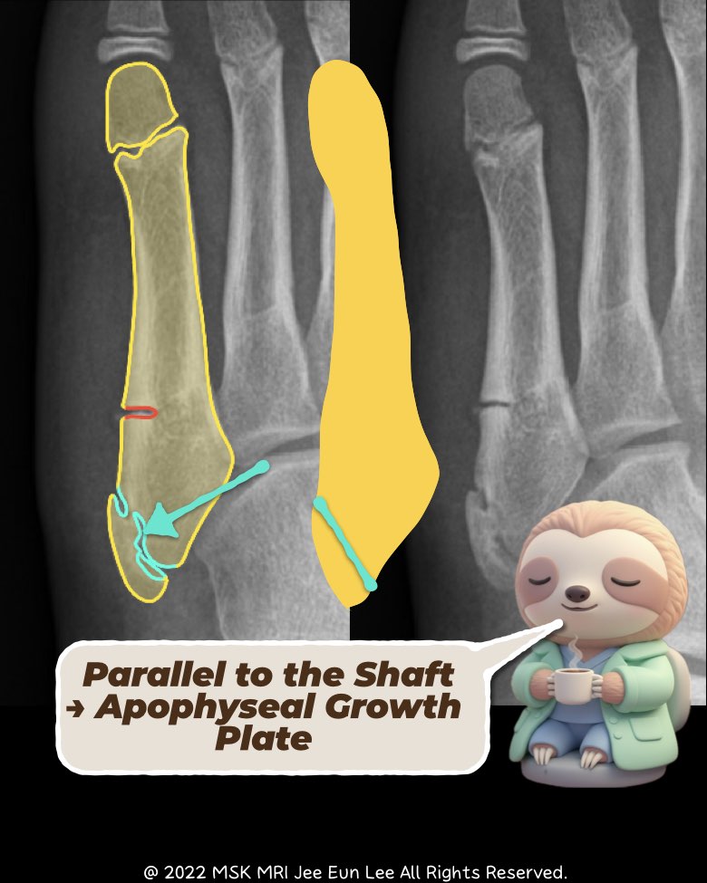
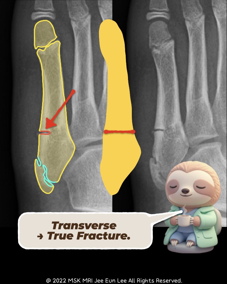
3. Radiographic Appearance
Apophysis
- Longitudinal/oblique radiolucent line.
- Parallel to the 5th metatarsal shaft.
- Does not extend into the cubometatarsal joint.
- Smooth margins reflecting orderly ossification.
Fracture
- Transverse or oblique line, orthogonal to shaft.
- Sharp, irregular edges.
- May extend into the articular surface (avulsion) or the metaphyseal–diaphyseal junction (Jones/stress fracture).
4. Symptoms and Physical Findings
Apophysis / Apophysitis
- Activity-related lateral foot pain.
- Point tenderness but minimal swelling and no ecchymosis.
- Radiographs may be normal or show mild irregularity.
Fracture
- Marked tenderness and visible swelling.
- Frequently includes ecchymosis.
- Pain causes difficulty with weight-bearing.
5. Course and Management
Apophysis (Normal Variant)
- No treatment required.
- Symptomatic Iselin disease responds to rest, activity modification, and NSAIDs.
- Apophysis fuses over 2–4 years.
Fracture
- Requires immobilization (boot or cast).
- Jones and diaphyseal stress fractures may require surgery due to nonunion risk.
- Correct classification is essential for prognosis.
6. Quick Comparison Table
| Feature | Apophysis (Normal/Apophysitis) | Fracture |
| Typical Age | 9–14 years | Any age |
| Trauma History | None or overuse | Clear acute trauma |
| Orientation | Parallel to shaft | Transverse/oblique |
| Joint Involvement | None | Possible |
| Soft-tissue Signs | Minimal swelling | Swelling, ecchymosis |
| Management | Observation / conservative | Immobilization ± surgery |
#5thmetatarsal, #apophysis, #IselinDisease, #footfracture, #radiologyeducation, #MSKradiology, #pediatricsportsmedicine, #fracturevsapophysis, #orthopedics, #musculoskeletalimaging, #Vibecase,
Visualizing MSK Radiology: A Practical Guide to Radiology Mastery
© 2022 MSK MRI Jee Eun Lee All Rights Reserved.
No unauthorized reproduction, redistribution, or use for AI training.
https://open.spotify.com/episode/2Zjh65CWfTgzsQaERYCCKl?si=5bFqkYOuSgytg1P8uFjlKw
📌 5th Metatarsal Base Fractures: Classification, Diagnosis, and Management
Dr. Slothic MSK Radiology Podcast · Episode
open.spotify.com