https://open.spotify.com/episode/2R70qYi5SeVcBIZhXuEY5i?si=NMnbnxJqRSSxFUQTBiyjoQ
A Radiologist’s Guide to Vascular Failure, Biomechanics, and Imaging Diagnosis
Why It Matters
Insufficiency fractures of the native patella after TKA are rare but frequently underdiagnosed.
These injuries arise from biomechanical overload applied to ischemic, metabolically weakened bone rather than acute trauma.
Radiologists are often the first to detect these subtle changes—even before symptoms become obvious.
1. Vascular Failure: The Seed of Insufficiency
Medial parapatellar arthrotomy disrupts the medial geniculate supply.
Lateral release may compromise the dominant superior lateral geniculate artery.
The combined effect leads to ischemia → osteonecrosis → microtrabecular failure → insufficiency fracture.
Imaging Indicators
- Patchy sclerosis
- Fragmented or “crumbling” patella
- Marrow signal changes disproportionate to symptoms
2. Biomechanics After TKA: A New Load Environment
The unresurfaced patella articulates directly with a metal trochlea, leading to:
- Point loading and stress-riser formation
- Significant increase in high-strain bone volume (up to ~200%)
- Elevated patellofemoral joint reaction forces (3–7× body weight)
Even routine activities may trigger stress remodeling → insufficiency fracture.
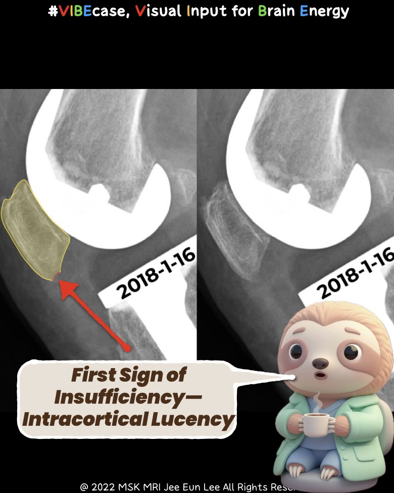
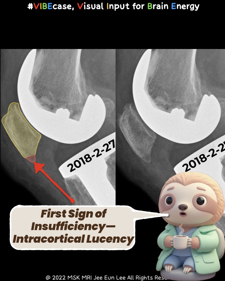
3. Radiography: Subtle but Crucial
Early Stage — Gray Cortex Sign
- Blurred anterior cortex
- Subtle intracortical lucency
- Reflects early osteoclastic tunneling
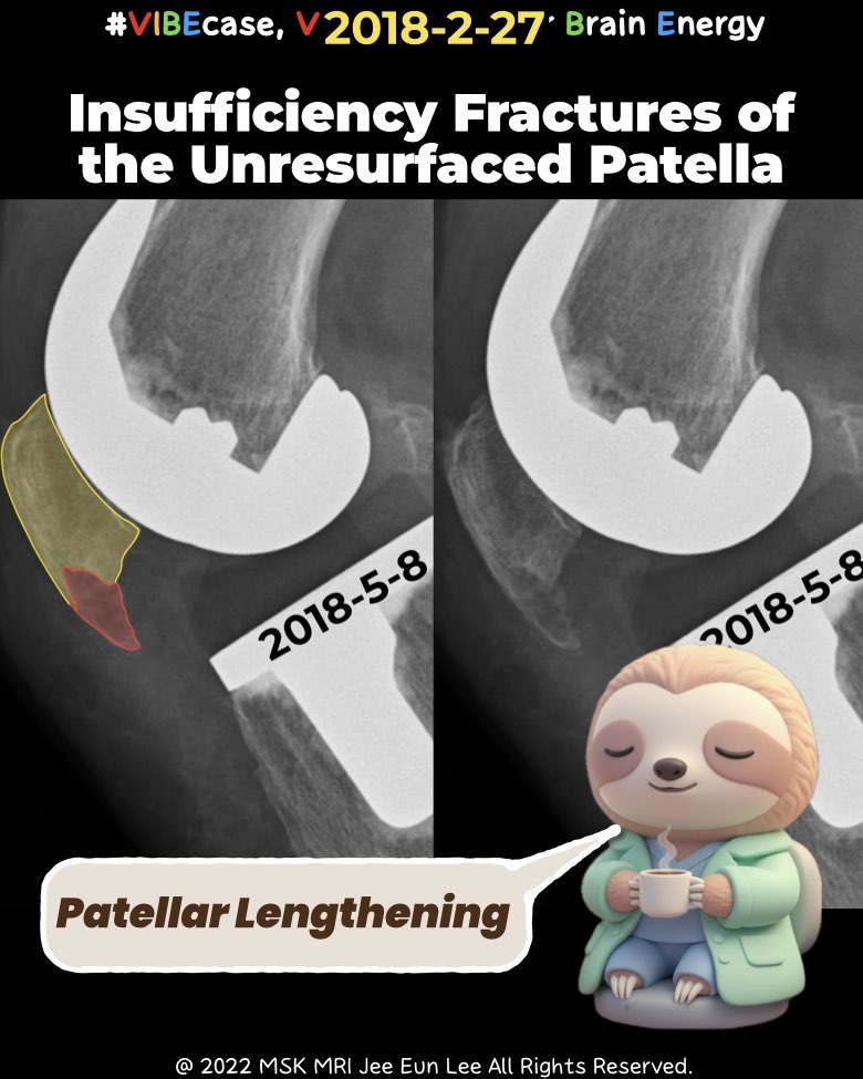
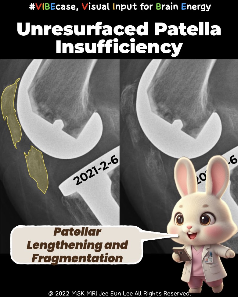
Late Stage
- Transverse fracture (most common)
- Vertical fracture (best seen on Skyline view)
- Marginal sclerosis indicating chronicity
- Patellar lengthening due to chronic distraction
- Fragmentation consistent with ischemic collapse
4. CT: Mapping What Radiographs Miss
- Detects occult vertical fractures
- Differentiates bipartite patella (smooth, corticated) from insufficiency fracture (irregular, cystic)
- Identifies subchondral cysts along the fracture plane
- Evaluates component rotation (internal rotation → lateral shear forces)
5. MRI: The Definitive Modality
Hallmark Finding
Diffuse bone marrow edema surrounding the fracture zone.
- T2/STIR: bright, extensive edema
- T1: linear low-signal fracture band
- Helps differentiate true fracture from bipartite patella (which lacks BME)
MRI provides the clearest depiction of the pre-fracture state, ischemic changes, and retinacular integrity.
Take-Home for Radiologists
- These injuries represent vascular and biomechanical failure, not traumatic disruption.
- Identify early imaging signs: gray cortex, subtle cortical blurring, marrow edema.
- MRI defines the diagnosis; CT clarifies morphology; SPECT assists when MRI is limited.
- Accurate early recognition prevents unnecessary intervention and supports effective conservative management.
#MSKRadiology, #TKAComplications, #PatellarFracture, #StressFractureMRI, #OrthopedicImaging,
#KneeMRI, #RadiologyEducation, #PeriprostheticFracture, #BoneMarrowEdema, #MusculoskeletalRadiology, #Vibecase
Visualizing MSK Radiology: A Practical Guide to Radiology Mastery
© 2022 MSK MRI Jee Eun Lee — All Rights Reserved.
Unauthorized reproduction, redistribution, or use for AI training is prohibited.