https://youtube.com/shorts/ykgTMN8cCG0
1. Why This Distinction Matters
The fifth metatarsal apophysis is a normal secondary ossification center in children and adolescents.
A fracture, in contrast, is a pathological break that follows trauma and usually requires treatment.
Key differences include age, orientation on imaging, clinical context, and soft-tissue findings.
2. Age and Clinical Context
Apophysis: Appears in skeletally immature patients (girls ~9–11, boys ~11–14). It is a normal developmental variant without clear trauma, sometimes presenting as overuse pain such as Iselin disease.
Fracture: Can occur at any age, especially in athletes. Typically follows an acute inversion injury with sudden pain, swelling, or ecchymosis.
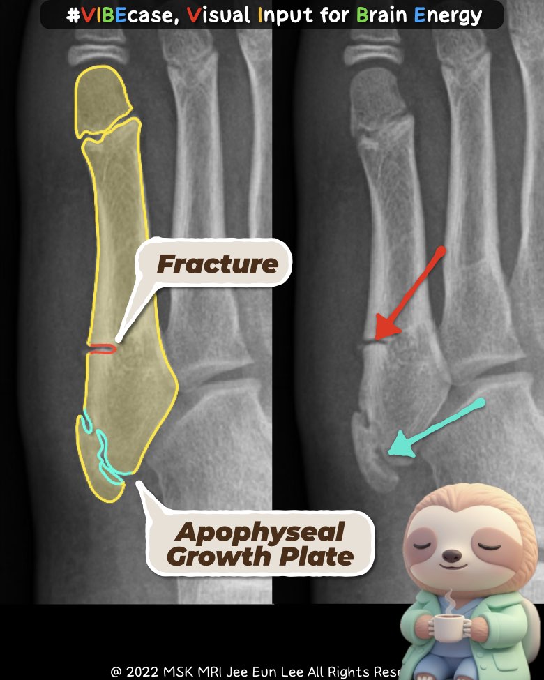
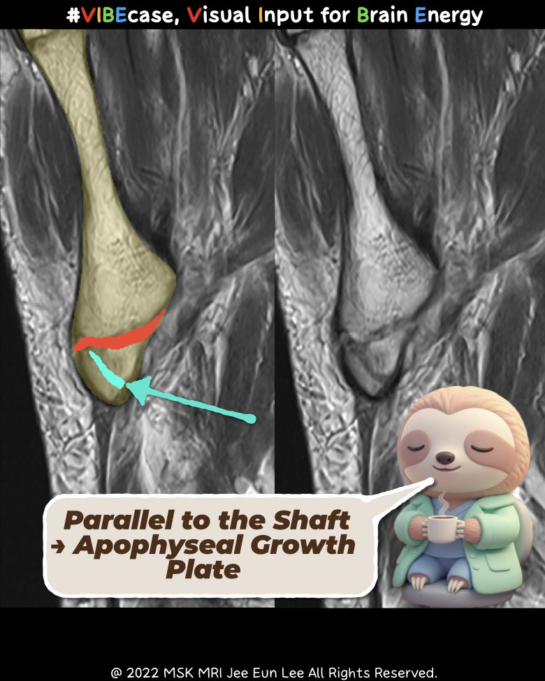
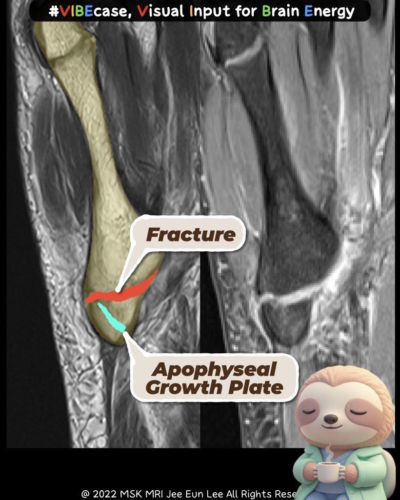
3. Radiographic Appearance
Apophysis: Shows a longitudinal or oblique radiolucent line running parallel to the 5th metatarsal shaft. Edges are smooth and it does not extend into the cubometatarsal joint.
Fracture: Appears as a transverse or oblique line that is orthogonal to the shaft. The margins are sharper and more irregular, and the line may extend into the articular surface, including avulsion or Jones/stress fracture patterns.
4. Symptoms and Physical Findings
Apophysis / Apophysitis: Causes activity-related lateral foot pain with point tenderness but usually minimal swelling and no ecchymosis. Radiographs may be normal or show mild irregularity.
Fracture: Presents with marked tenderness, visible swelling, and often ecchymosis. Weight-bearing becomes painful or difficult.
5. Course and Management
Apophysis (Normal Variant): No treatment is required. Symptomatic Iselin disease improves with rest, activity modification, and NSAIDs, and the apophysis naturally fuses over 2–4 years.
Fracture: Requires immobilization with a boot or cast. Jones and diaphyseal stress fractures carry a higher nonunion risk and may need surgical intervention. Accurate classification is essential.
#5thmetatarsal, #apophysis, #IselinDisease, #footfracture, #radiologyeducation, #MSKradiology, #pediatricsportsmedicine, #fracturevsapophysis, #orthopedics, #musculoskeletalimaging, #Vibecase
Visualizing MSK Radiology: A Practical Guide to Radiology Mastery
© 2022 MSK MRI Jee Eun Lee All Rights Reserved.
No unauthorized reproduction, redistribution, or use for AI training.
https://open.spotify.com/episode/2Zjh65CWfTgzsQaERYCCKl?si=5bFqkYOuSgytg1P8uFjlKw
📌 5th Metatarsal Base Fractures: Classification, Diagnosis, and Management
Dr. Slothic MSK Radiology Podcast · Episode
open.spotify.com
https://open.spotify.com/episode/7wJqd534Vmqx8y6KBSkxwB?si=4hz_MM9ESk2H2WMWN593lQ
[Lecture] 📌 Understanding the Difference: Fifth Metatarsal Apophysis vs. Fracture
Dr. Slothic MSK Radiology Podcast · Episode
open.spotify.com