https://youtube.com/shorts/gBfJcQGh48I
Concept
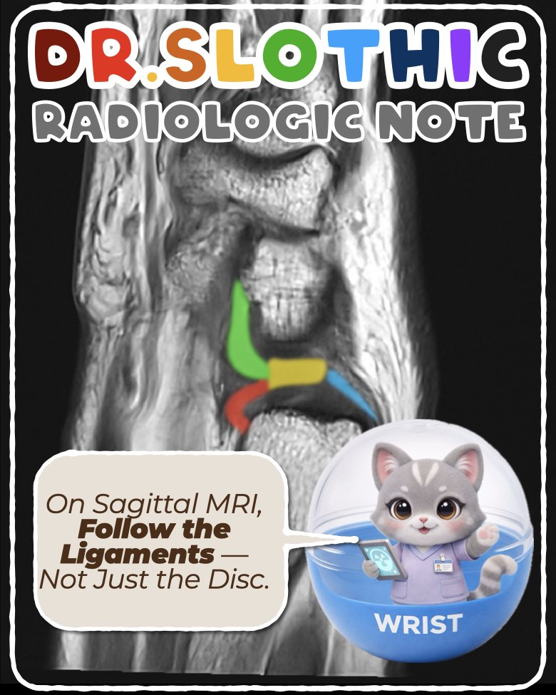
On sagittal wrist magnetic resonance imaging,
the triangular fibrocartilage complex should not be read as a single disc.
It represents a continuous ulnar-sided stabilizing system connecting the distal radioulnar joint to the ulnocarpal joint.
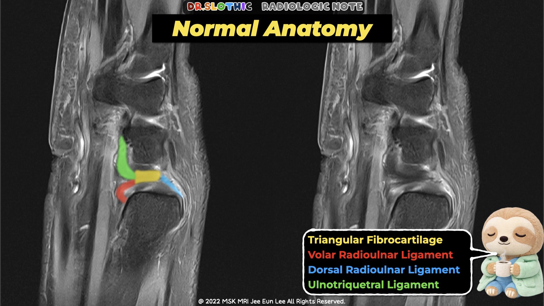
Central Anchor — Articular Disc
- The articular disc lies at the center of the triangular fibrocartilage complex.
- Its dorsal and volar margins are not free edges.
- They blend directly into the dorsal and volar radioulnar ligaments, forming the core stabilizing arms.
Radioulnar Ligaments
Dorsal Radioulnar Ligament
- Thickened dorsal peripheral margin of the articular disc.
- Extends from the dorsal rim of the distal radius (sigmoid notch)
to the ulnar styloid and fovea of the ulnar head. - Consists of:
- Becomes taut in pronation and provides dorsal stability of the distal radioulnar joint.
- Blends dorsally with the floor of the extensor carpi ulnaris tendon sheath.
Volar Radioulnar Ligament
- Thickened volar peripheral margin of the articular disc.
- Originates from the palmar rim of the sigmoid notch and lunate facet of the distal radius.
- Also composed of superficial (styloid) and deep (foveal) fibers.
- Tightens in supination, acting as the primary volar stabilizer of the distal radioulnar joint.
- Serves as the proximal origin for the major palmar ulnocarpal ligaments.
Ulnocarpal Ligaments (Distal Continuation)
Ulnolunate Ligament
- Palmar ligament extending from the volar radioulnar ligament region
and indirectly from the ulnar fovea or styloid base. - Inserts on the palmar cortex of the lunate.
- Courses distally along the long axis of the forearm.
- Helps suspend the lunate and resist palmar and ulnar translation of the proximal carpal row.
Ulnotriquetral Ligament
- Palmar ligament arising mainly from the volar radioulnar ligament,
with partial contribution from the palmar-ulnar aspect of the ulnar styloid. - Fans out distally to the proximal and ulnar surfaces of the triquetrum.
- Lies just ulnar to the ulnolunate ligament.
- Contributes to ulnocarpal stability and load transmission from the ulnar carpus to the ulna.
MRI Take-Home Message
On sagittal wrist magnetic resonance imaging:
disc → radioulnar ligaments → ulnocarpal ligaments
should be followed as one continuous stabilizing chain.
Instability rarely comes from the disc alone.
#DrSlothic #SlothicRadiology #MSKMRI #WristMRI #SagittalMRI #WristAnatomy #TFCC #UlnarSidedWrist #DistalRadioulnarJoint #UlnocarpalLigaments #RadiologyEducation #MSKRadiology #HandWristMRI
Visualizing MSK Radiology: A Practical Guide to Radiology Mastery
© 2022 MSK MRI Jee Eun Lee All Rights Reserved.
No unauthorized reproduction, redistribution, or use for AI training.


'✅ Dr. Slothic Notes' 카테고리의 다른 글
| Shoulder MRI in Real Practice: From Rotator Cuff to Instability | Core Cases (0) | 2026.02.20 |
|---|---|
| 📌 Galeazzi Fracture–Dislocation — Wrist MRI Focus (0) | 2026.02.01 |
| 📌 Galeazzi Fracture–Dislocation (0) | 2026.02.01 |
| 📌 The Most Common Calf Tear Pattern — Don’t Miss the Transverse Tear (0) | 2026.01.28 |
| 📌 Why This Is NOT a Strain — Muscle and Nerve Tell the Story (0) | 2026.01.27 |