https://youtube.com/shorts/FB--s6N052w
Key Radiologic Points
Clinical Context
- Sudden calf pain during uphill running
- Sharp pulling sensation
- Classic presentation of acute calf strain
Where to Start on MRI
- Always begin with axial images
- In calf injuries, vertical tears are the diagnostic starting point, not a secondary finding
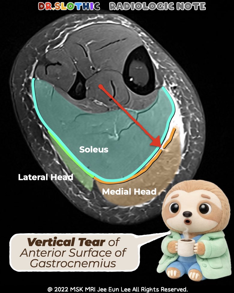
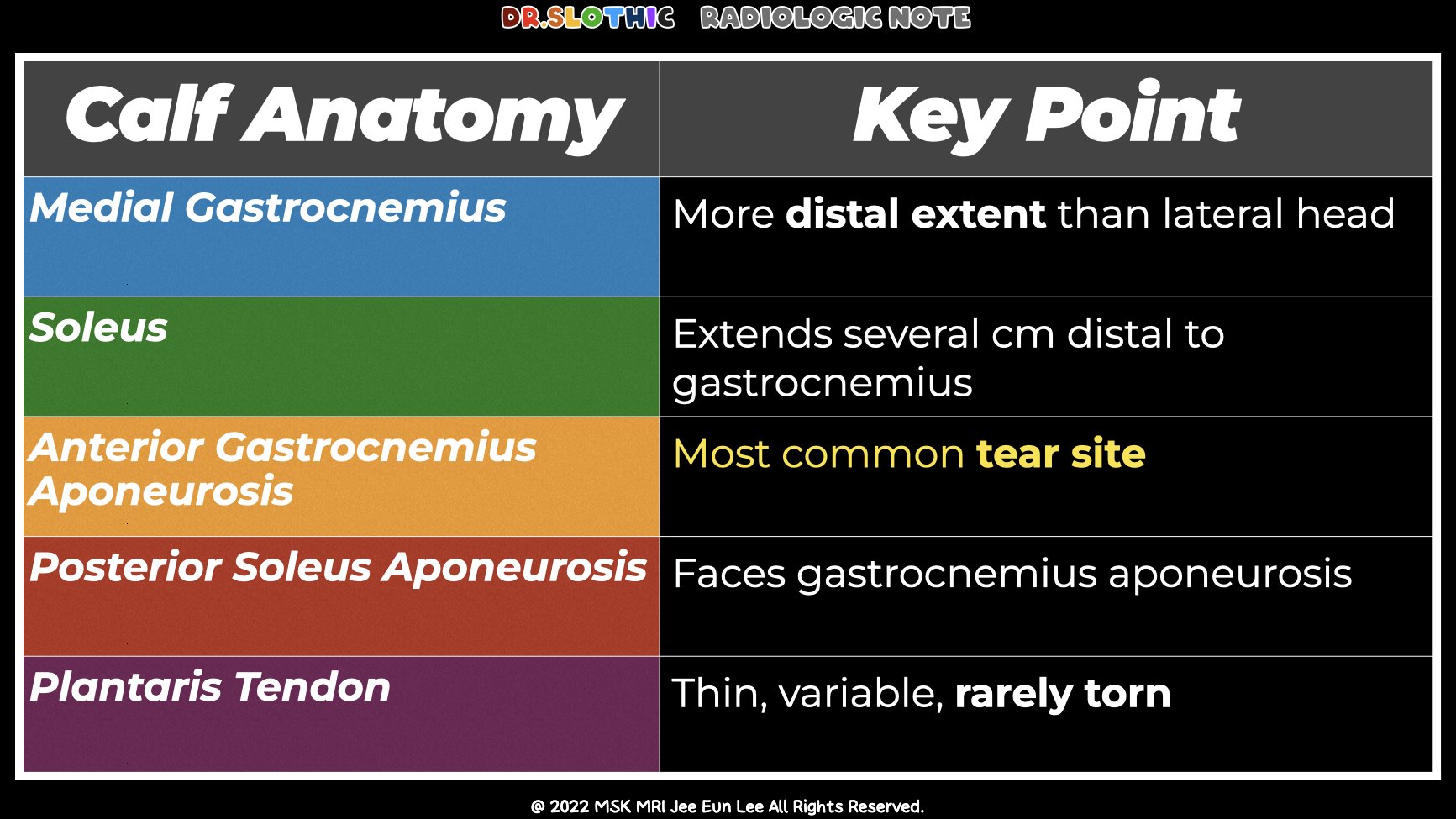
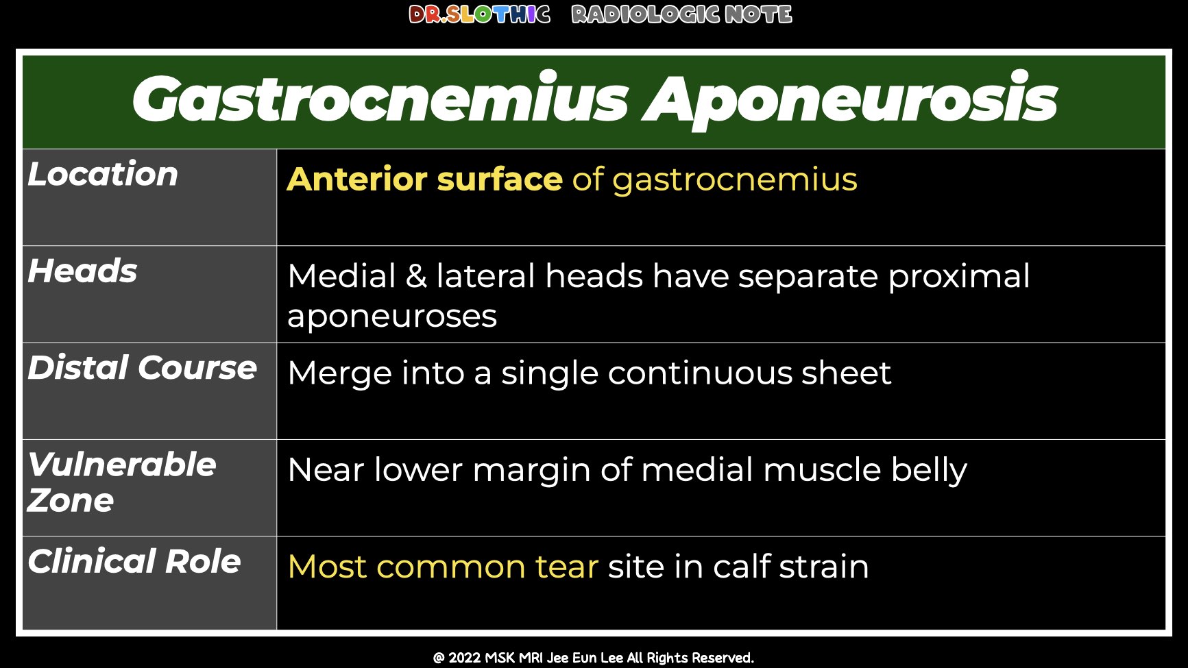
Key Anatomic Landmark
- Focus on the anterior aponeurosis of the medial gastrocnemius
- Normally seen as a thin, smooth, arcing low-signal line along the anterior muscle surface
Vertical Tear
- Focal interruption of the anterior aponeurosis
- Best found by inspecting areas adjacent to muscle edema or hematoma
- Once identified, it must be followed slice by slice
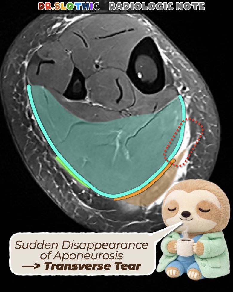
How to Find the Transverse Tear
- Track the same aponeurosis inferiorly
- Look for the sudden disappearance of the aponeurosis
- Muscle is present, but the aponeurosis is missing
- This combination defines a transverse tear gap
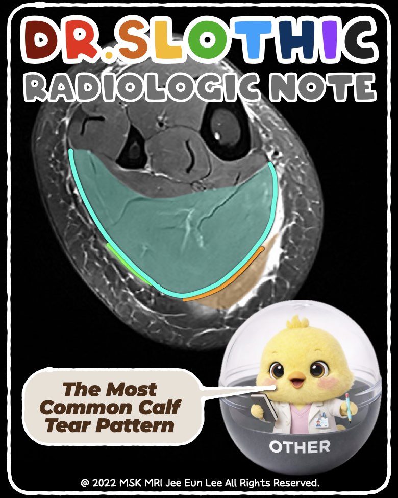
Most Common Pattern: L-Pattern Tear
- Primary partial-width transverse tear of the medial gastrocnemius aponeurosis
- Associated vertical tear at the lateral margin of the detached segment
- Inferior transverse component: usually full-thickness
- Vertical and superior components: typically partial-thickness
Important Pitfall
- Mild soleus edema may be present
- No injury to intramuscular tendons or aponeurotic junctions
- Recognize it, but do not let it distract from the primary pattern
Take-Home Message
- Vertical tears are the entry point to diagnosis
- Always follow them inferiorly to identify the transverse component
- Learn the L-pattern tear — it is the most common calf tear pattern
- Pattern recognition makes the diagnosis straightforward
#DrSlothic #SlothicRadiology #MSKMRI #CalfTear #CalfStrain #Gastrocnemius
#SportsMRI #MuscleInjury #PatternRecognition #RadiologyEducation
Visualizing MSK Radiology: A Practical Guide to Radiology Mastery
© 2022 MSK MRI Jee Eun Lee All Rights Reserved.
No unauthorized reproduction, redistribution, or use for AI training.


'✅ Dr. Slothic Notes' 카테고리의 다른 글
| 📌 Galeazzi Fracture–Dislocation — Wrist MRI Focus (0) | 2026.02.01 |
|---|---|
| 📌 Galeazzi Fracture–Dislocation (0) | 2026.02.01 |
| 📌 Why This Is NOT a Strain — Muscle and Nerve Tell the Story (0) | 2026.01.27 |
| 📌 When Elbow Edema Isn’t a Tear — MRI Clues to Delayed onset muscle soreness (0) | 2026.01.26 |
| 📌 Localized Tenosynovial Giant Cell Tumor of the Knee (0) | 2026.01.24 |