https://youtube.com/shorts/aqm32avcGcA
Radiologic Essentials
1. Start With the Pattern
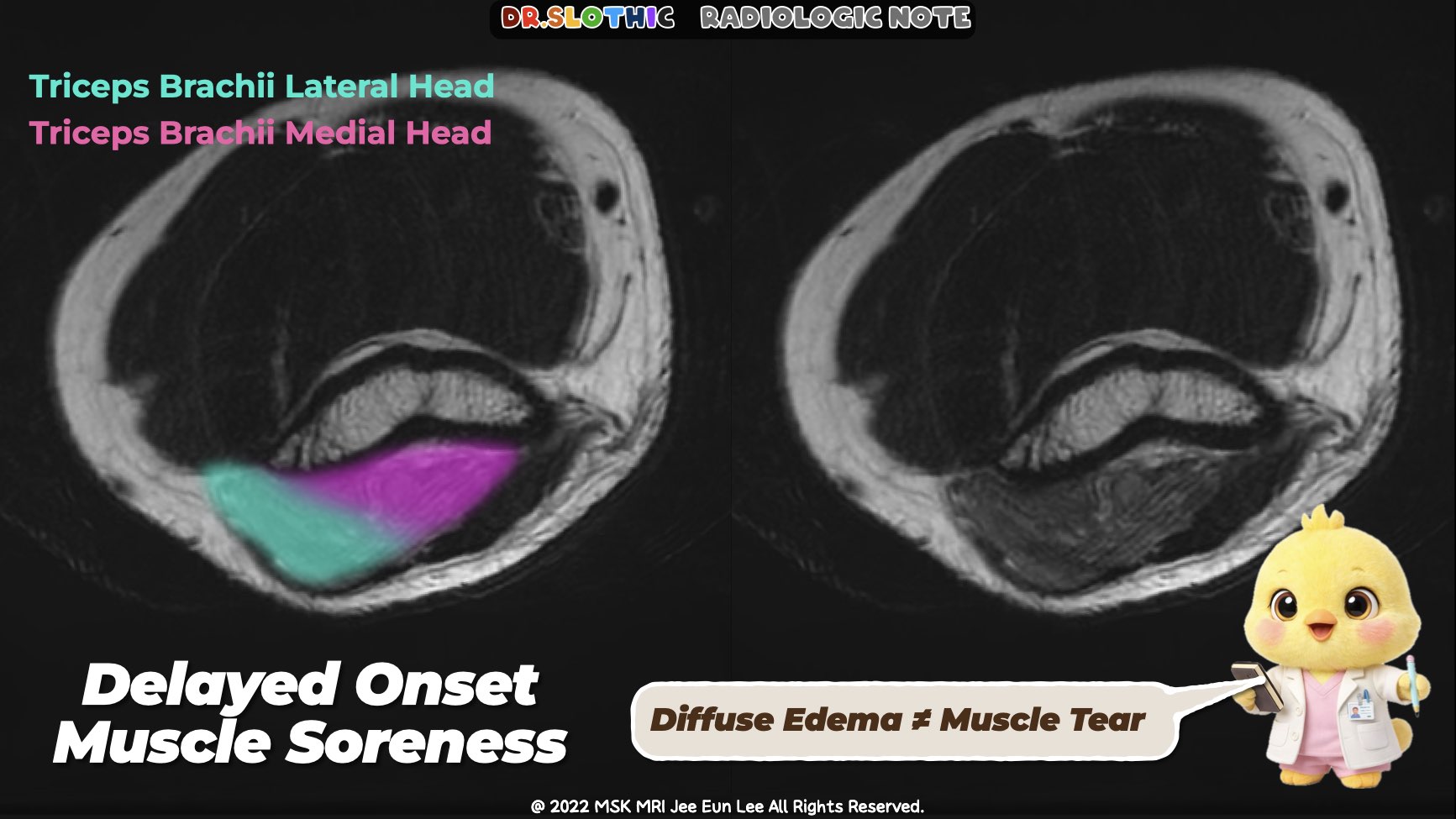
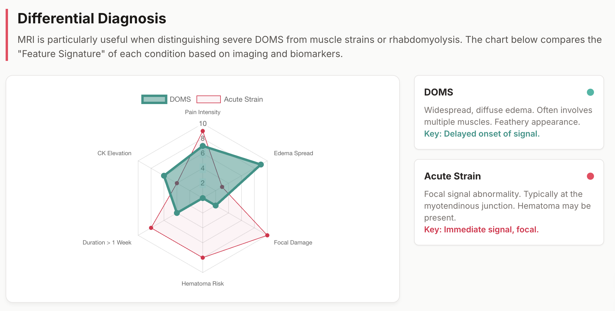
• Fluid-sensitive sequences show diffuse or patchy high signal
• Typically involves multiple muscles within a functional group
• This distribution immediately argues against a focal traumatic tear
2. Architecture Is the Key Anchor
• Fascicular architecture remains intact
• No macroscopic fiber disruption, retraction, or large hematoma
• Muscle bulk may be mildly increased but remains organized
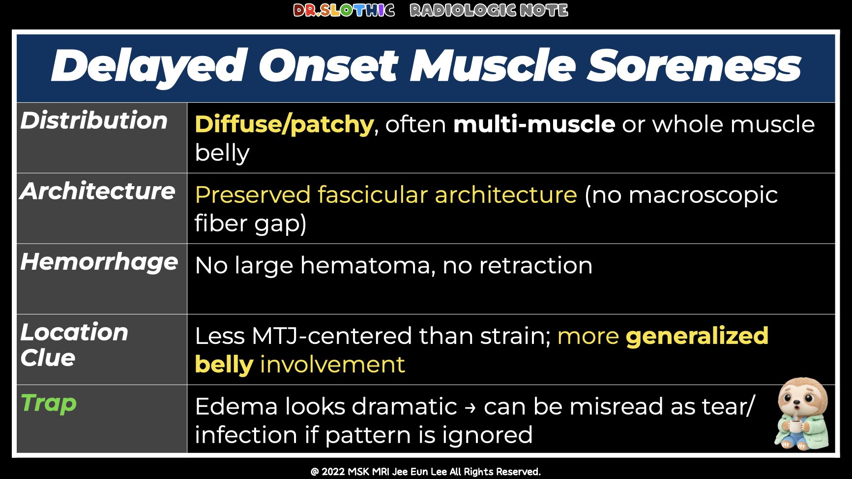
3. Location Helps Exclude Strain
• Signal abnormality is not MTJ-centered
• More generalized involvement of the muscle belly
• Unlike Grade 1 strain, no feathery MTJ-tracking edema
4. Don’t Ignore the Fascia
• Deep fascial edema is commonly present
• Fascia has high nociceptor density and acts as a major pain generator
• Explains pain that appears disproportionate to structural findings
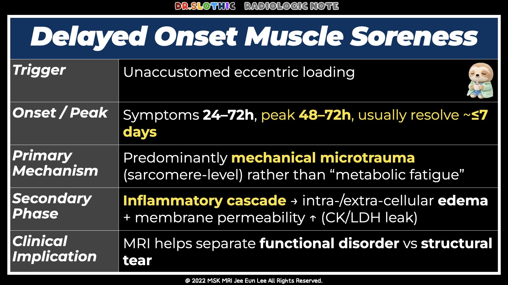
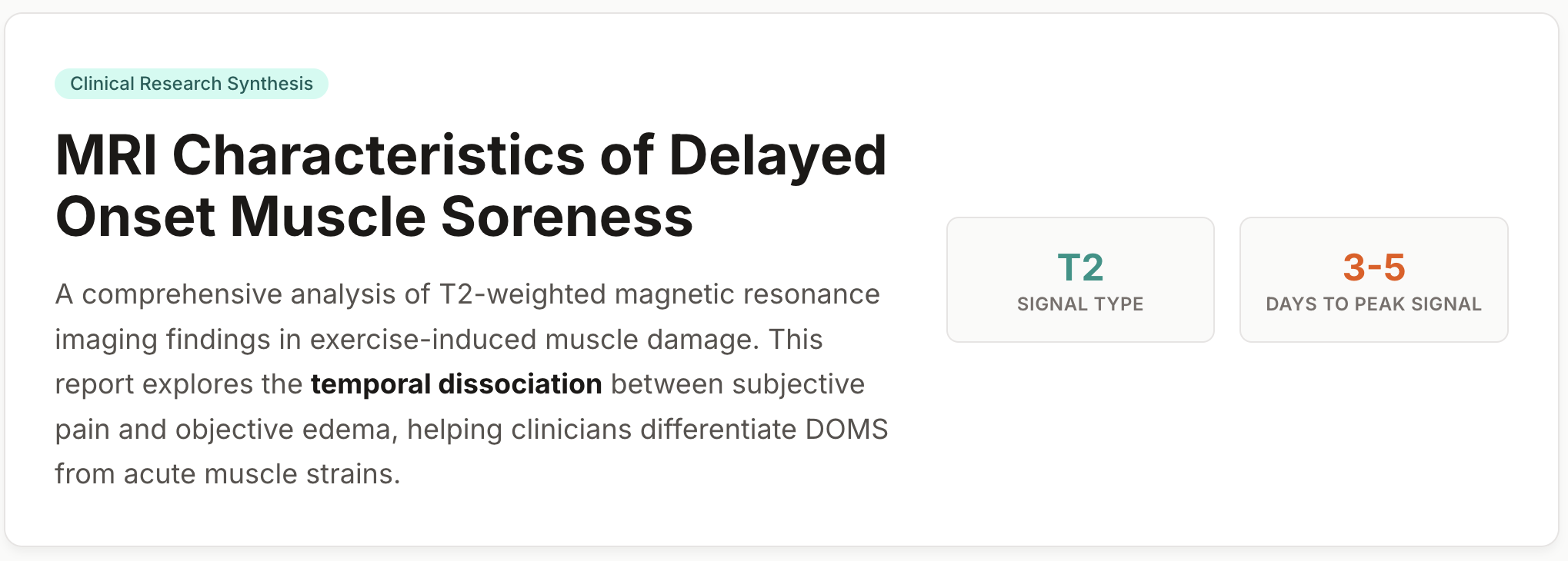
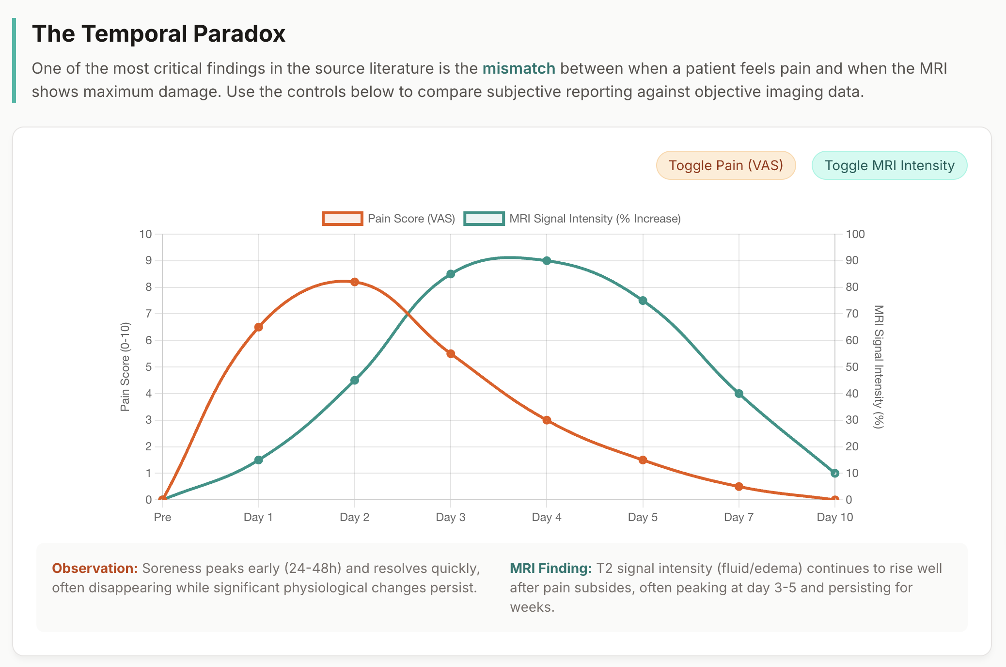
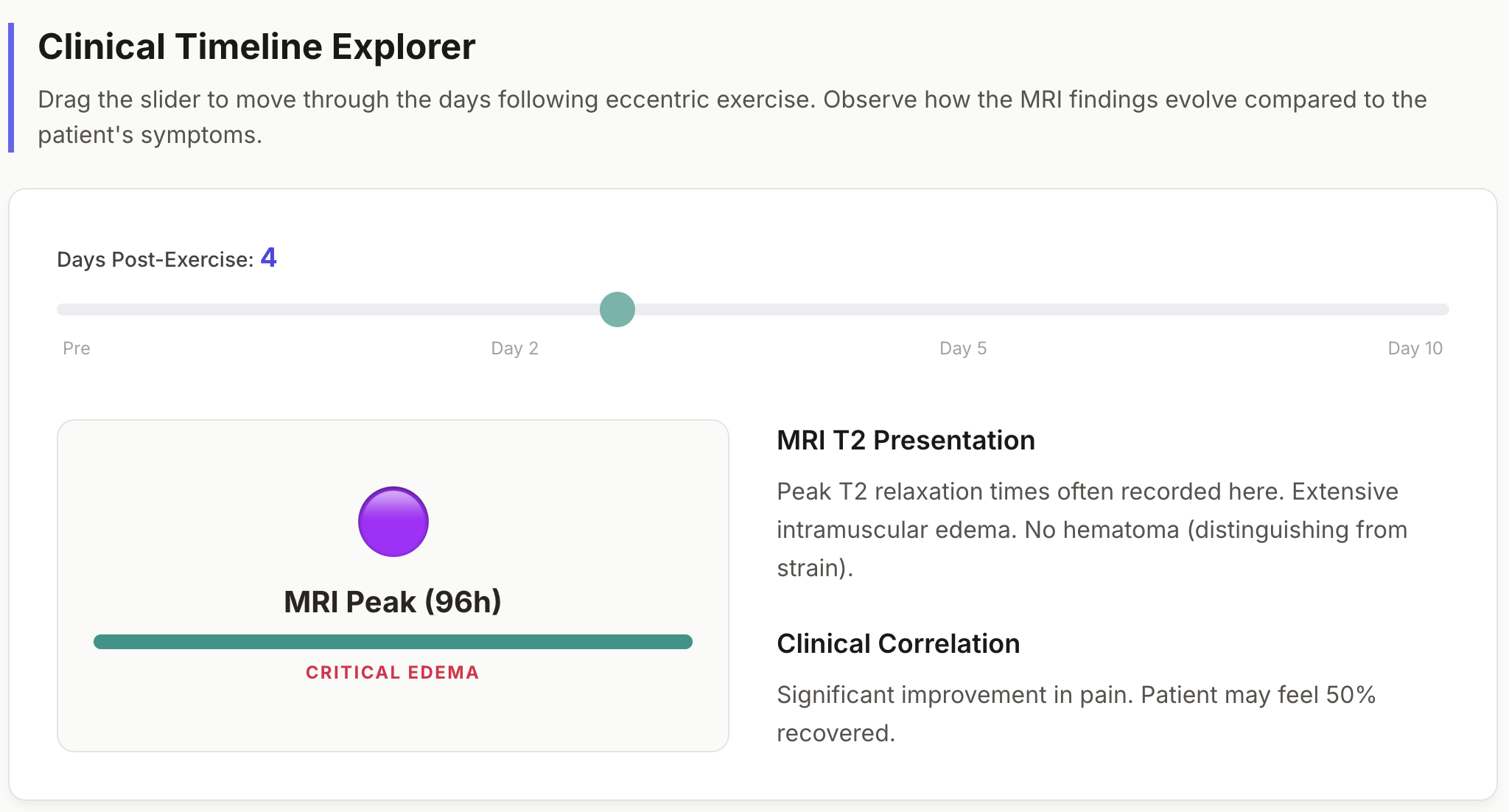
5. Expect MRI–Clinical Dissociation
• STIR/T2 hyperintensity may persist weeks to months
• Imaging abnormalities often outlast symptom resolution
• Persistent edema does not equal ongoing injury
Radiologic Take-Home Message
Diffuse, reversible muscle ± fascial edema
with preserved internal architecture
→ favors Delayed Onset Muscle Soreness (DOMS)
→ a functional muscle disorder, not a structural tear
#SportsMRI #DOMS #MuscleEdema #ElbowMRI #MSKRadiology
#SportsImaging #PatternRecognition #RadiologyEducation #DrSlothic #SlothicRadiology #MSKMRI
Visualizing MSK Radiology: A Practical Guide to Radiology Mastery
© 2022 MSK MRI Jee Eun Lee All Rights Reserved.
No unauthorized reproduction, redistribution, or use for AI training.


'✅ Dr. Slothic Notes' 카테고리의 다른 글
| 📌 The Most Common Calf Tear Pattern — Don’t Miss the Transverse Tear (0) | 2026.01.28 |
|---|---|
| 📌 Why This Is NOT a Strain — Muscle and Nerve Tell the Story (0) | 2026.01.27 |
| 📌 Localized Tenosynovial Giant Cell Tumor of the Knee (0) | 2026.01.24 |
| 📌 Cyclops Lesion After ACL Reconstruction (0) | 2026.01.24 |
| 📌 Bennett Lesion and Posterosuperior Internal Impingement: A Common Pitfall (0) | 2026.01.20 |