Muscle edema doesn’t always mean strain.
These cases represent crush injury.
Read them like strain, and you will miss what matters.
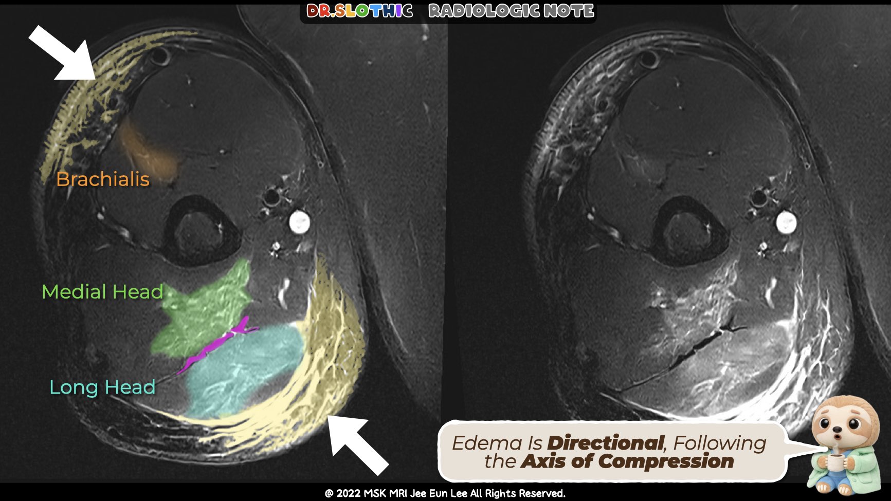
Case 1 — Muscle Crush Injury (Triceps-dominant)
- Mechanism: Direct industrial anterior–posterior compression of the upper arm
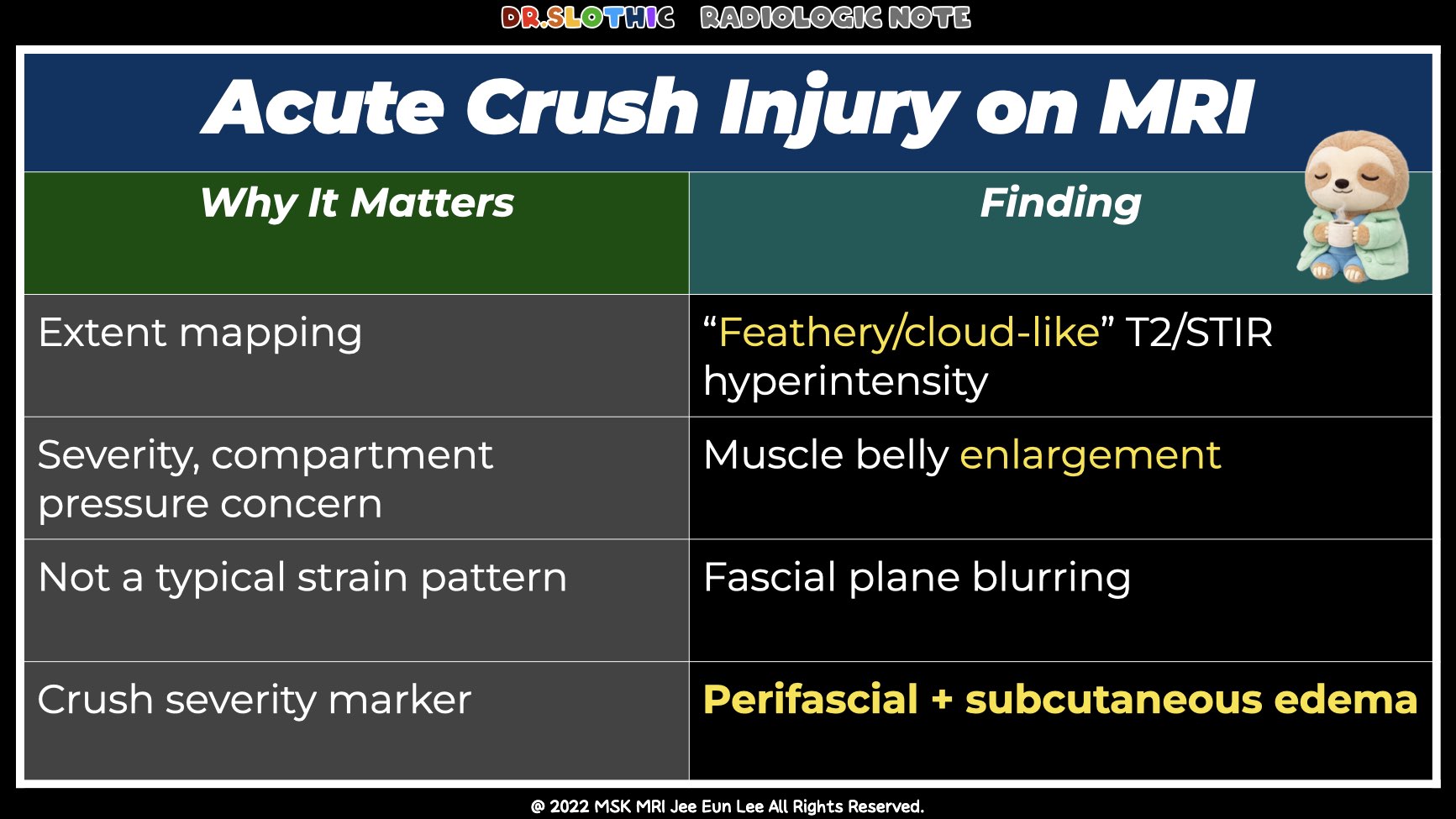
- MRI (fat-suppressed T2):
- Peripheral fascial fluid tracking and perifascial edema present
- Myotendinous junction relatively spared
- Dominant involvement of the muscle belly, not the tendon
- Subtle brachialis involvement with adjacent subcutaneous edema
→ Muscle compressed between bone and subcutaneous layer
Interpretation:
Compression-related muscle belly injury → true muscle crush injury, not strain or DOMS.
Case 2 — Radial Nerve Involvement in Crush Injury
- Clinical: Arm numbness and finger extension weakness, no humeral fracture
- Anatomy: Radial nerve vulnerable at the spiral groove, directly against bone
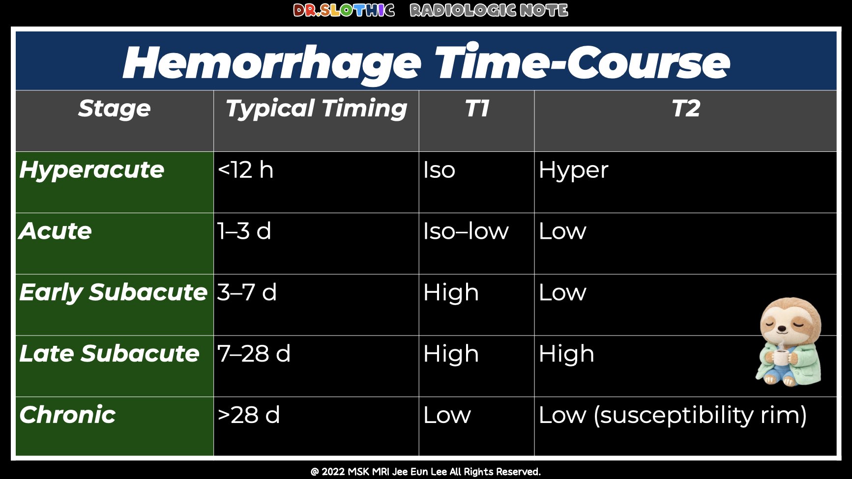
- MRI findings:
- Associated brachialis intramuscular hematoma and muscle edema
Interpretation:
Nerve injury occurs within the same global crush pattern
→ Compression-related neuropathy (neurapraxia to axonotmesis)
Key reporting phrase:
“No definite nerve transection is identified.”
Final Take-Home Message
- Crush injury is a distinct trauma entity
- Muscle clues:
- Nerve clues:
- Always interpret muscle and nerve together, guided by mechanism and anatomy
#CrushInjury #MuscleCrush #TraumaticMyopathy #CompressionInjury #RadialNerveInjury #MSKMRI #MuscleEdema #NotAStrain #RadiologyEducation #PatternRecognition #DrSlothic #SlothicRadiology #MSKMRI
Visualizing MSK Radiology: A Practical Guide to Radiology Mastery
© 2022 MSK MRI Jee Eun Lee All Rights Reserved.
No unauthorized reproduction, redistribution, or use for AI training.


'✅ Dr. Slothic Notes' 카테고리의 다른 글
| 📌 Galeazzi Fracture–Dislocation (0) | 2026.02.01 |
|---|---|
| 📌 The Most Common Calf Tear Pattern — Don’t Miss the Transverse Tear (0) | 2026.01.28 |
| 📌 When Elbow Edema Isn’t a Tear — MRI Clues to Delayed onset muscle soreness (0) | 2026.01.26 |
| 📌 Localized Tenosynovial Giant Cell Tumor of the Knee (0) | 2026.01.24 |
| 📌 Cyclops Lesion After ACL Reconstruction (0) | 2026.01.24 |