https://youtube.com/shorts/X5KaN_j65m8
A Practical MRI Guide for Radiologists
Cyclops lesion, or localized anterior arthrofibrosis, is one of the most common and most easily overcalled findings after ACL reconstruction.
Because it is familiar, it often becomes a reflex diagnosis.
This is exactly why understanding its typical timing, location, and behavior matters.
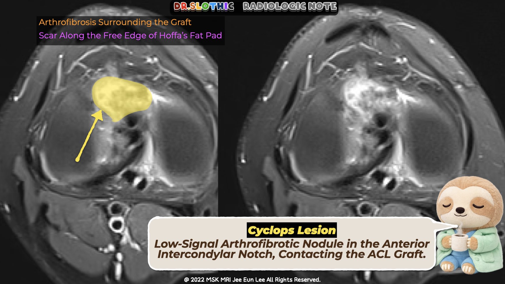
What a True Cyclops Lesion Looks Like
Clinical context
- Develops early, typically within the first postoperative year
- Often associated with loss of terminal extension, sometimes with a “cyclops clunk”
Typical MRI location
- Anterior intercondylar notch
- Immediately anterior to the tibial insertion of the ACL graft
- Often gently displaces the posterior margin of Hoffa’s fat pad
- Does not track along synovium
Morphology
- Small, well-circumscribed nodular mass
- Ovoid or bulbous
- Stable in size over time
Signal characteristics
- T1-weighted: low to intermediate signal
- T2 / PD fat-sat: heterogeneous, but not markedly low
- STIR: relatively hyperintense (granulation component)
- GRE / SWI: no susceptibility blooming
Why Cyclops Lesions Form
Cyclops lesions represent a localized fibroproliferative response related to:
- Surgical debris from tibial tunnel drilling
- Residual ACL stump tissue
- Early graft impingement
- Postoperative inflammatory milieu
Importantly, they form early and typically do not continue to grow over years.
When You Should Stop Calling It Cyclops
Be cautious if an anterior notch lesion:
- Appears many years after surgery
- Progressively enlarges
- Becomes lobulated
- Evolves to predominantly low T2 signal
- Shows hemorrhagic signal or GRE blooming
That pattern is not typical for postoperative arthrofibrosis and should prompt consideration of synovial-based pathology, particularly localized tenosynovial giant cell tumor.
Take-Home Message
Cyclops lesion is not just a location-based diagnosis.
Timeline, stability, and signal behavior matter.
Early, small, stable, anterior to the tibial ACL insertion — that is cyclops.
Growth and signal evolution over time mean you should rethink the diagnosis.
#CyclopsLesion #ACLReconstruction #KneeMRI #MSKRadiology #PostoperativeMRI #AnteriorIntercondylarNotch #Arthrofibrosis #RadiologyEducation #DrSlothic #SlothicRadiology #DrSlothic #SlothicRadiology
Visualizing MSK Radiology: A Practical Guide to Radiology Mastery
© 2022 MSK MRI Jee Eun Lee All Rights Reserved.
No unauthorized reproduction, redistribution, or use for AI training.


'✅ Dr. Slothic Notes' 카테고리의 다른 글
| 📌 When Elbow Edema Isn’t a Tear — MRI Clues to Delayed onset muscle soreness (0) | 2026.01.26 |
|---|---|
| 📌 Localized Tenosynovial Giant Cell Tumor of the Knee (0) | 2026.01.24 |
| 📌 Bennett Lesion and Posterosuperior Internal Impingement: A Common Pitfall (0) | 2026.01.20 |
| 📌 Labral Calcification: The MRI Low-Signal Trap (0) | 2026.01.18 |
| 📌 Unstable OCD in Adults: What MRI Is Really Telling You (0) | 2026.01.18 |