https://youtube.com/shorts/BKlONA0UWag
https://youtube.com/shorts/6yHDS7sore4
📍 Watch Related Shorts
📌 Bennett Lesion: What It Really Represents on Shoulder CT
👉 https://youtube.com/shorts/BKlONA0UWag
📌 Labral Calcification: The MRI Low-Signal Trap
👉 https://youtube.com/shorts/_UNZ5r3dH1M
📌 Hill-Sachs fracture??— Same CT Look, Different Mechanism
👉 https://youtube.com/shorts/6yHDS7sore4
Let’s look at a case and ask two simple questions.
Case Overview
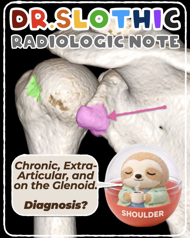
On radiographs and CT:
- Extra-articular ossification / exostosis along the glenoid rim
- Irregular, elongated, crescent-shaped bony density
- Well-corticated margins → chronic process
- Adjacent glenoid rim sclerosis
- Not a loose body
Clinical context:
- Long-term amateur baseball pitcher
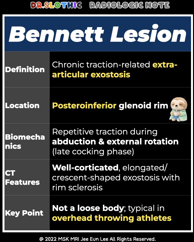
→ Diagnosis: Bennett lesion
Bennett lesion is a chronic traction-related exostosis arising from the posteroinferior glenoid rim,
representing traction enthesopathy at the posterior capsulolabral attachment,
related to repetitive stress in abduction and external rotation, especially the late cocking phase.
Question 1 — Same Area, Different Diagnosis
Another patient shows:
- Amorphous, high-attenuation intra-articular calcification
- Localized to the posterosuperior labrum
- Bulky, partially fragmented deposit
- MRI: marked edema and labral contour loss
Clinical context:
- 59-year-old woman
- Acute severe shoulder pain for 4 days
→ This is not a Bennett lesion.
→ Basic Calcium Phosphate deposition of the labrum (acute calcific labritis).
Question 2 — Hill-Sachs Fracture?
In the Bennett lesion case:
- Subtle posterior humeral head flattening is present
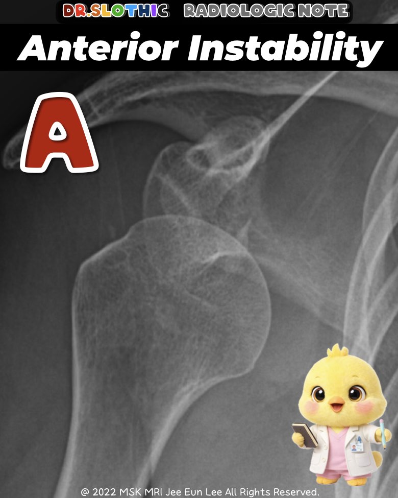
Compare with a true anterior instability case:
- Bony Bankart lesion
- Medially displaced fragment
- Classic Hill-Sachs fracture
Back to the Bennett lesion patient:
- No anterior instability
- No bony Bankart lesion
→ Not a Hill-Sachs fracture
So What Is It?
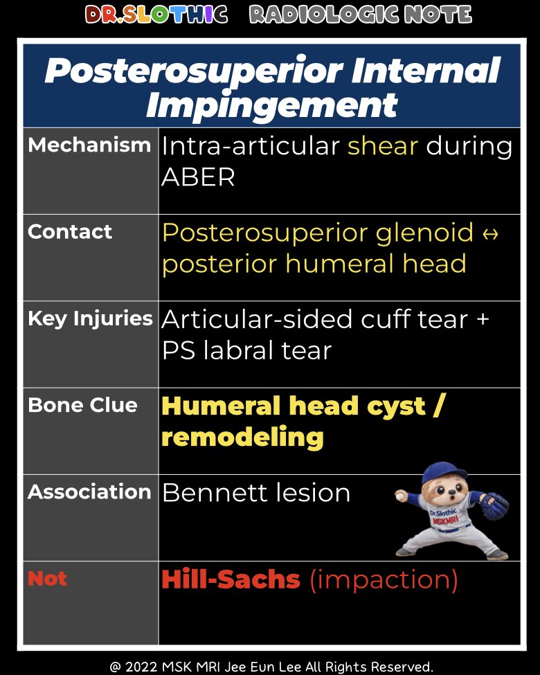
This represents posterosuperior internal impingement.
- In abduction and external rotation,
the glenohumeral contact point shifts posterosuperiorly - Leads to repetitive shear stress
- Affects the undersurface of the rotator cuff and posterosuperior labrum
Typical MRI Findings
- Posterior/posterosuperior humeral head cysts
- Articular-sided partial-thickness tears
- Undersurface fraying, not bursal-sided
- Posterosuperior labral tear
- Often associated with Bennett lesion or posterior capsular tightness
Take-Home Message
If a CT looks like a Hill-Sachs fracture but the anterior instability pattern is absent,
think chronic remodeling from posterosuperior internal impingement, not a fracture.
#DrSlothic #SlothicRadiology #BennettLesion #HillSachs #InternalImpingement #ShoulderCT #ShoulderMRI #SportsRadiology #MSKRadiology #RadiologyPitfalls
Visualizing MSK Radiology: A Practical Guide to Radiology Mastery
© 2022 MSK MRI Jee Eun Lee All Rights Reserved.
No unauthorized reproduction, redistribution, or use for AI training.


'✅ Dr. Slothic Notes' 카테고리의 다른 글
| 📌 Localized Tenosynovial Giant Cell Tumor of the Knee (0) | 2026.01.24 |
|---|---|
| 📌 Cyclops Lesion After ACL Reconstruction (0) | 2026.01.24 |
| 📌 Labral Calcification: The MRI Low-Signal Trap (0) | 2026.01.18 |
| 📌 Unstable OCD in Adults: What MRI Is Really Telling You (0) | 2026.01.18 |
| 📌 The Gravid Hip: Subchondral Insufficiency Fracture of the Femoral Head (Pregnancy & Puerperium) (0) | 2026.01.18 |