https://youtube.com/shorts/_UNZ5r3dH1M
Calcific Labritis (BCP Deposition in the Glenoid Labrum)
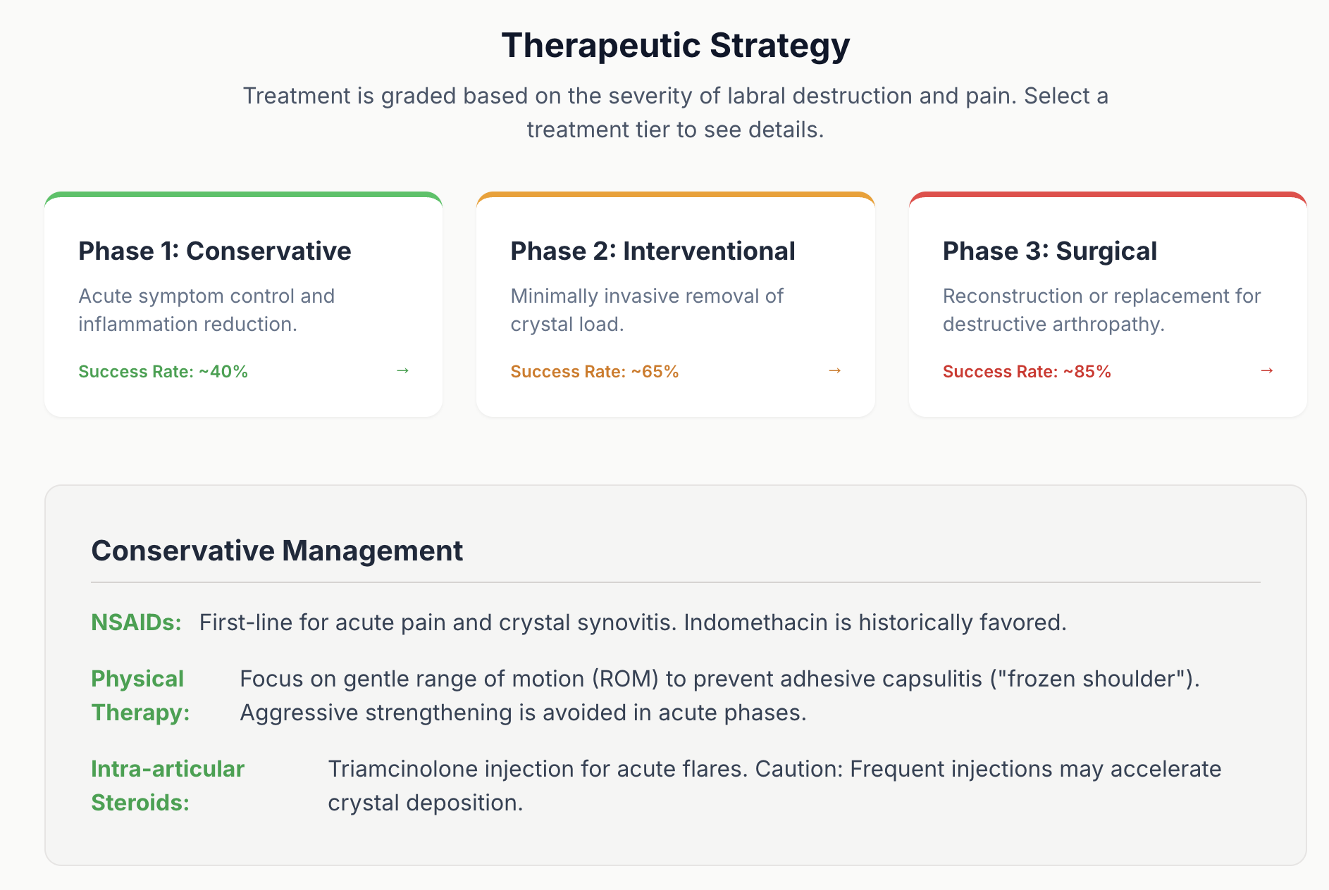
Why this matters
- Shoulder calcific tendinitis is common
- Labral calcification is rare
- Rare things get miscalled
- The common miscalls are expensive: infection or labral tear
What it is
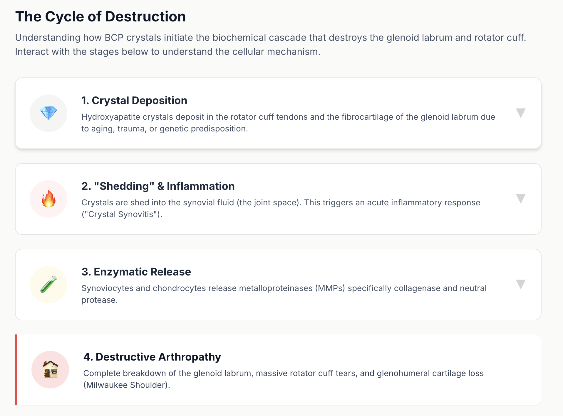
- Basic Calcium Phosphate (BCP) crystal deposition
- Hydroxyapatite-type calcium
- Can deposit inside the glenohumeral joint
- Can be embedded in the glenoid labrum
- The painful presentation is often the acute inflammatory (resorptive) phase
Why pain can be extreme
- This is crystal-induced chemical synovitis
- Pain is often sudden
- Pain is often out of proportion
- ROM shuts down due to inflammation, not weakness
Imaging logic (the only workflow that works)
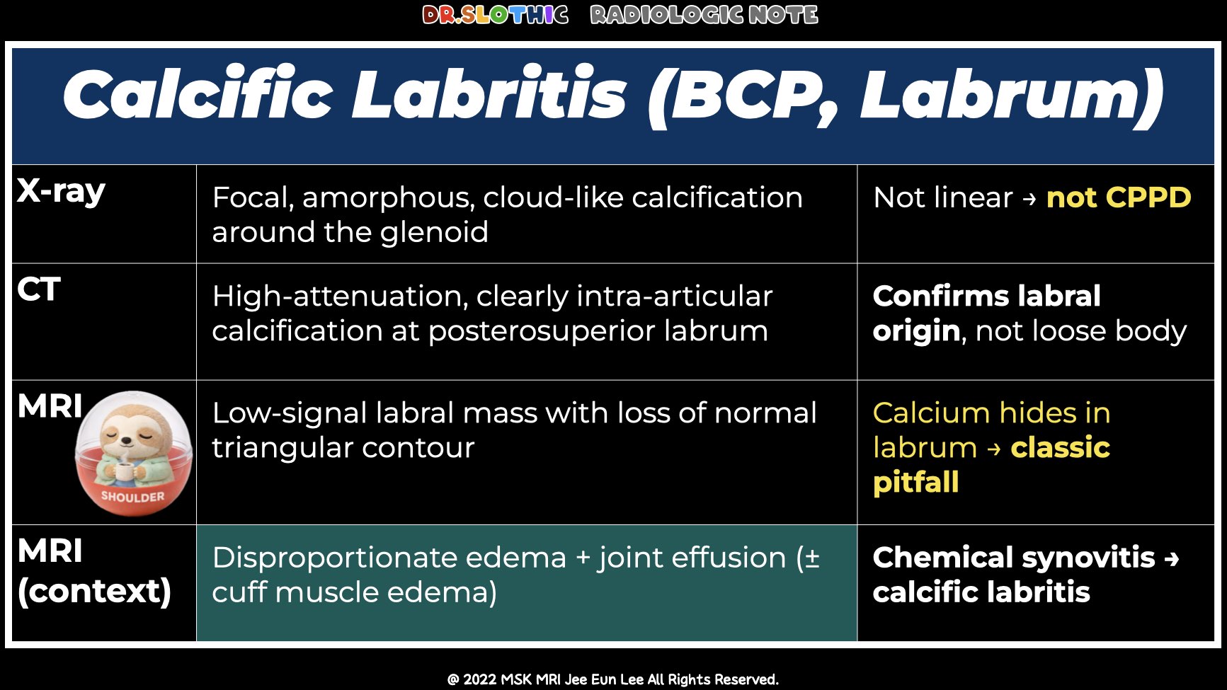
1) X-ray: detect the “type”
- BCP tends to look dense / amorphous / cloud-like
- CPPD tends to look thin / linear / punctate
- The diagnostic question becomes: where is it?
2) CT: prove the “location”
- Best modality to confirm intra-articular calcification
- Best modality to localize to labrum / biceps anchor / glenoid rim
- Also prevents the classic mistake: calling it a loose body or fracture
3) MRI: don’t chase calcium
- Calcium is low signal on essentially all sequences
- The labrum is low signal too
- So the deposit can “disappear” into the labrum
MRI diagnosis is pattern-based:
- Loss of normal labral contour
- Amorphous low-signal focus replacing the labral triangle
- Disproportionate inflammation
- Edema can make the lesion more conspicuous
- But edema also triggers misdiagnosis if you ignore X-ray/CT
Arthroscopy (what it explains)
- Hyperemic, inflamed labrum
- Incision can release chalky / toothpaste-like material
- Evacuation removes the inflammatory driver
- Repair depends on residual stability, not on the presence of calcium
Take-Home Message
- X-ray detects the suspicious calcification pattern
- CT localizes it to the labrum and confirms it is intra-articular
- MRI misleads unless you look for contour loss + disproportionate inflammation
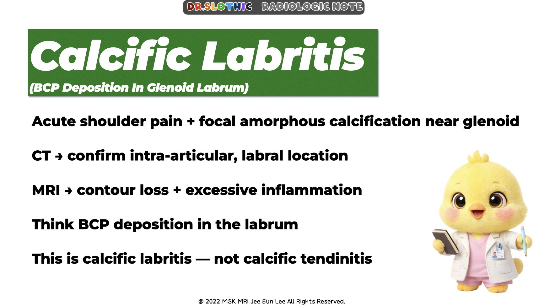
If acute shoulder pain shows:
- focal amorphous calcification near the glenoid
- CT-proven intra-articular labral location
- MRI with labral contour loss and marked inflammatory reaction
Think: BCP deposition in the glenoid labrum — calcific labritis.
Call it early, and you avoid labeling it as infection or a confusing labral tear.
#DrSlothic #SlothicRadiology #MSKMRI #CalcificLabritis #BCP #Hydroxyapatite #ShoulderMRI #GlenoidLabrum #CrystalArthropathy #RadiologyPearls #MSKRadiology #OrthopedicImaging #CTCorrelation #XrayFirst #DontChaseEdema
Visualizing MSK Radiology: A Practical Guide to Radiology Mastery
© 2022 MSK MRI Jee Eun Lee All Rights Reserved.
No unauthorized reproduction, redistribution, or use for AI training.


'✅ Dr. Slothic Notes' 카테고리의 다른 글
| 📌 Cyclops Lesion After ACL Reconstruction (0) | 2026.01.24 |
|---|---|
| 📌 Bennett Lesion and Posterosuperior Internal Impingement: A Common Pitfall (0) | 2026.01.20 |
| 📌 Unstable OCD in Adults: What MRI Is Really Telling You (0) | 2026.01.18 |
| 📌 The Gravid Hip: Subchondral Insufficiency Fracture of the Femoral Head (Pregnancy & Puerperium) (0) | 2026.01.18 |
| 📌 Radial Nerve Palsy After Crush Injury (0) | 2026.01.17 |