https://youtube.com/shorts/OtO-fqnDCpc
MRI as the Diagnostic Gold Standard
MRI is the key modality for diagnosing localized TGCT (L-TGCT), defining extent, and planning surgery.
Its multiplanar capability and soft-tissue contrast reliably differentiate L-TGCT from other intra-articular masses.
Key MRI Findings (Imaging-Focused)
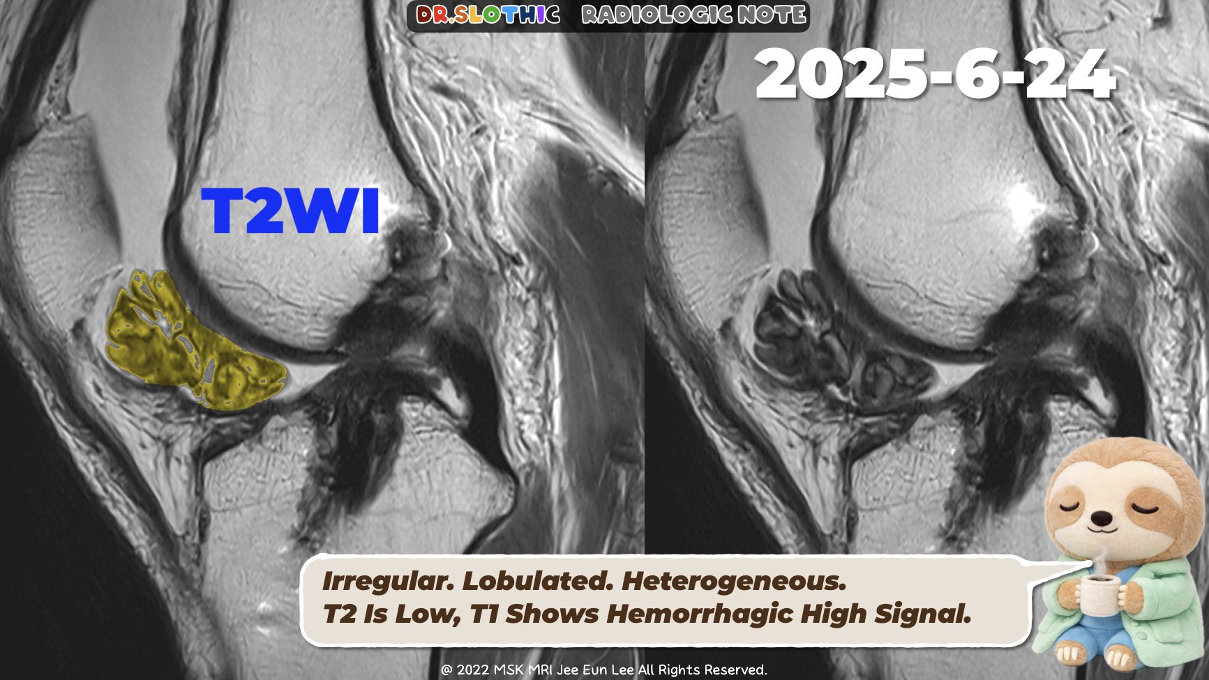
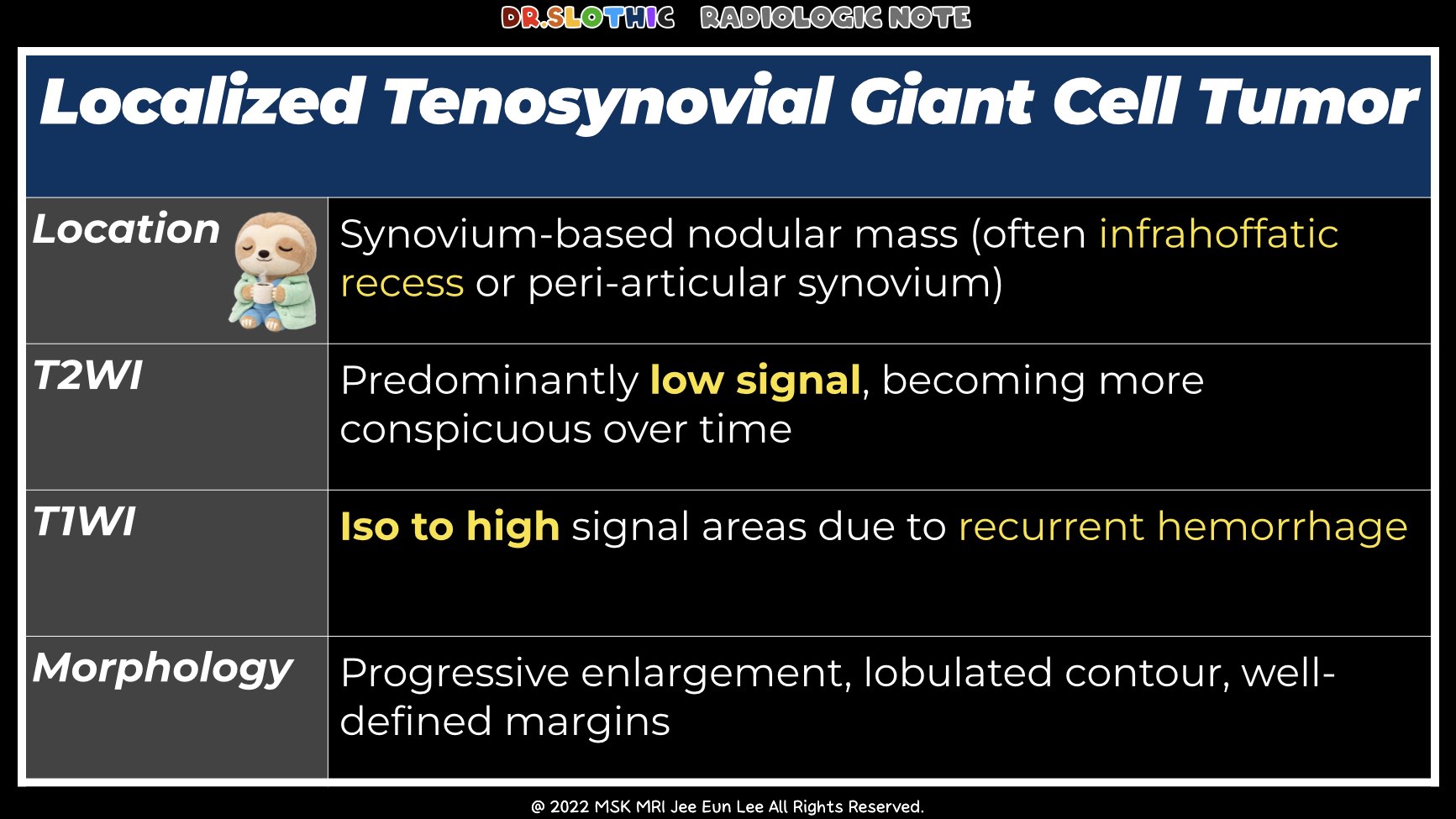
Morphology
- Solitary, well-circumscribed, lobulated nodular mass
- Distinct from diffuse TGCT (no carpet-like synovial infiltration)
- Often encapsulated by a thin low-signal pseudocapsule
T1-Weighted Imaging
- Low to intermediate signal, usually iso–slightly hypointense to muscle
- Helps exclude lipoma and simple subacute hematoma
T2 / Fluid-Sensitive Imaging
- Heterogeneous signal
- Occasional cleft-like hyperintensity between lobules
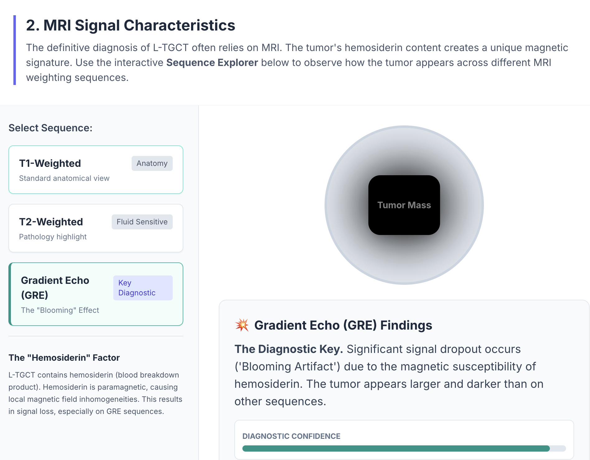
GRE / SWI
- Blooming artifact from hemosiderin (may be subtle in localized type)
- Absence of marked blooming does not exclude L-TGCT
Post-Contrast (T1 C+)
- Moderate to intense, heterogeneous enhancement
- Differentiates from ganglion cyst (rim only) and hematoma (no internal enhancement)
Key Differentials (MRI Clues)
- Fibroma of tendon sheath: very low T1/T2, no blooming, less heterogeneous enhancement
- Synovial hemangioma: very high T2, serpentine vessels, phleboliths
- Synovial sarcoma: T2 triple signal, calcification, aggressive growth
- Synovial chondromatosis: multiple cartilaginous nodules, calcification/ossification
- Hoffa’s disease: diffuse fat-pad edema or fibrosis, no discrete blooming nodule
Take-Home
A synovium-based nodular mass with low T2 signal, (variable) blooming on GRE, and heterogeneous enhancement strongly favors localized TGCT, especially when morphology and evolution do not fit postoperative fibrosis.
Hashtags
#LocalizedTGCT #TenosynovialGiantCellTumor #KneeMRI #MSKRadiology #IntraArticularMass
#T2LowSignal #GREBlooming #SynovialTumor #DifferentialDiagnosis #RadiologyEducation #DrSlothic #SlothicRadiology
Visualizing MSK Radiology: A Practical Guide to Radiology Mastery
© 2022 MSK MRI Jee Eun Lee All Rights Reserved.
No unauthorized reproduction, redistribution, or use for AI training.


'✅ Dr. Slothic Notes' 카테고리의 다른 글
| 📌 Why This Is NOT a Strain — Muscle and Nerve Tell the Story (0) | 2026.01.27 |
|---|---|
| 📌 When Elbow Edema Isn’t a Tear — MRI Clues to Delayed onset muscle soreness (0) | 2026.01.26 |
| 📌 Cyclops Lesion After ACL Reconstruction (0) | 2026.01.24 |
| 📌 Bennett Lesion and Posterosuperior Internal Impingement: A Common Pitfall (0) | 2026.01.20 |
| 📌 Labral Calcification: The MRI Low-Signal Trap (0) | 2026.01.18 |