https://youtube.com/shorts/WipVHnQZo3g
Why the wrist matters
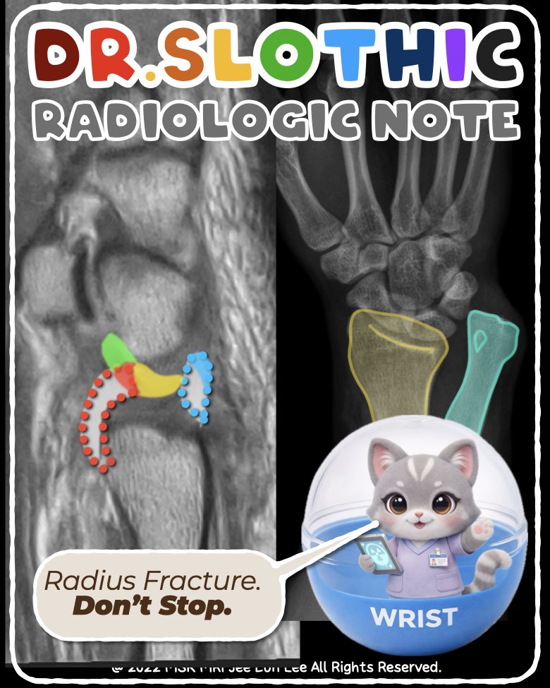
- Galeazzi fracture is not only a radial shaft injury.
- The key problem is often DRUJ instability, caused by disruption of the TFCC stabilizers.
Sagittal MRI: Best plane for ulnar-sided stabilizers
- Sagittal imaging clearly demonstrates the TFCC ligamentous components, not just the articular disc.
Normal TFCC Stabilizing Core
- The central articular disc is present,
but true DRUJ stability depends on the radioulnar ligaments:
Dorsal Radioulnar Ligament
- Dorsal ulnar head → distal radius
- Tight in pronation
Volar Radioulnar Ligament
- Volar ulnar head → distal radius
- Tight in supination
➡️ Tears of these ligaments lead directly to DRUJ instability.
Distal Continuation: Volar Ulnocarpal Sling
- The volar radioulnar ligament continues into:
These form the volar ulnocarpal stabilizing sling.
MRI Findings in This Galeazzi Case
- Ulnar styloid process fracture
- ECU tendon longitudinal splitting
- Combined tears of:
Take-home Message
This is not just a fracture.
It is a complete disruption of the ulnar-sided stabilizers, explaining persistent DRUJ instability.
#DrSlothic #SlothicRadiology #MSKMRI #GaleazziFracture #GaleazziFractureDislocation #DistalRadioulnarJoint #DRUJ #ForearmTrauma #OrthopedicRadiology #TraumaRadiology #MSKRadiology #RadiologyEducation #RadiologyPearls #CaseBasedRadiology #OrthopedicImaging #XrayInterpretation #CTinTrauma #PostoperativeImaging
Visualizing MSK Radiology: A Practical Guide to Radiology Mastery
© 2022 MSK MRI Jee Eun Lee All Rights Reserved.
No unauthorized reproduction, redistribution, or use for AI training.


'✅ Dr. Slothic Notes' 카테고리의 다른 글
| 📌 Sagittal Wrist MRI Anatomy_Ulnar-Sided Stabilizing System (0) | 2026.02.01 |
|---|---|
| 📌 Galeazzi Fracture–Dislocation (0) | 2026.02.01 |
| 📌 The Most Common Calf Tear Pattern — Don’t Miss the Transverse Tear (0) | 2026.01.28 |
| 📌 Why This Is NOT a Strain — Muscle and Nerve Tell the Story (0) | 2026.01.27 |
| 📌 When Elbow Edema Isn’t a Tear — MRI Clues to Delayed onset muscle soreness (0) | 2026.01.26 |