ATFL The ATFL is best imaged on axial T1 or high-resolution PD MR images, appearing as a flat, thin, homogeneous band of low signal intensity arising from the anterior margin of the lateral malleolus and coursing anteromedially downward to attach onto the neck of the talus, just anterior to the fibular articular cartilage.
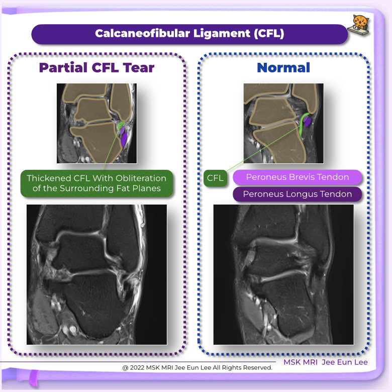
The CFL is large, strong, and cordlike. It arises from the deep aspect of the inferior tip of the lateral malleolus and courses posteroinferiorly to attach to the lateral aspect of the calcaneus, just about the retro-trochlear eminence. The CFL crosses both the tibiotalar and subtalar joints and is located deep to the peroneal tendons.
🥰 #followme,
https://www.instagram.com/msk_mri/
💟 Visit my youtube channel for a detailed description
Youtube channel: MSKMRI
#AnkleMRI, #ATFL, #CFL, #ATFLtear, #CFLtear, #Ankleligament, #ligamenttear,
#안산에이스병원,#AnsanAceHospital, #MSKMRI, #mskmri_Ankle, #영상의학과이지은, #영상의학공부맛집, #studygram, #MRI자신감키우기, #군자출판사,