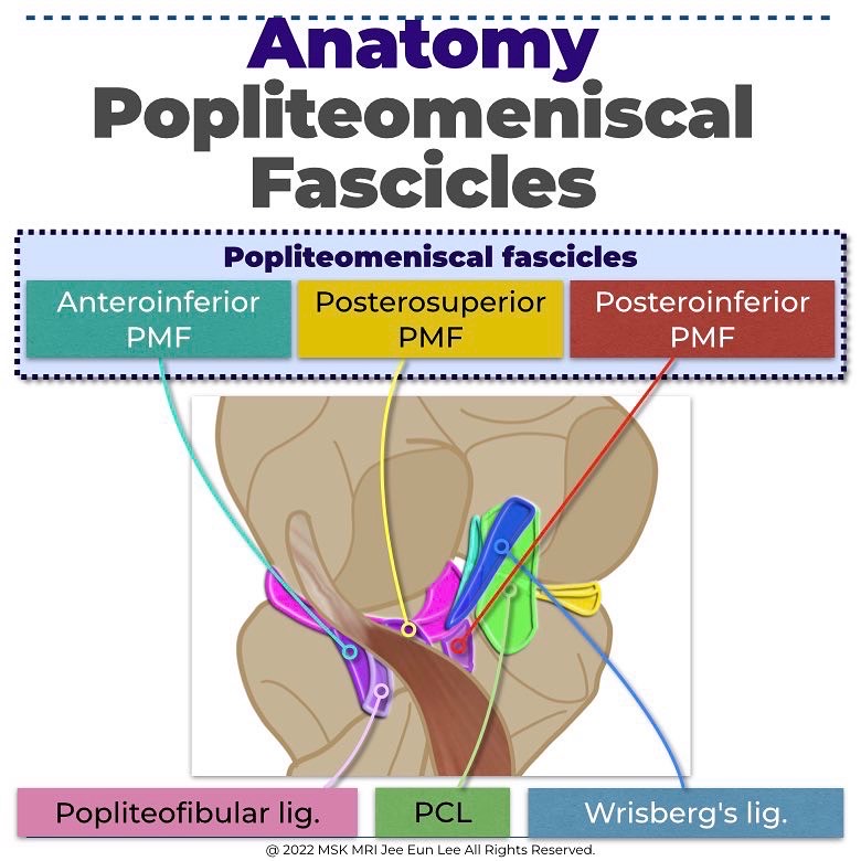
* The popliteomeniscal fascicles arise from the periphery of the posterior horn of the lateral meniscus and form a posterolateral meniscocapsular extension, which creates the popliteus hiatus.
* The fascicles not only form the hiatus, which allows the popliteus tendon an avenue to course intra-articularly and maintain the integrity of the joint but stabilize and control the motion of the posterior horn
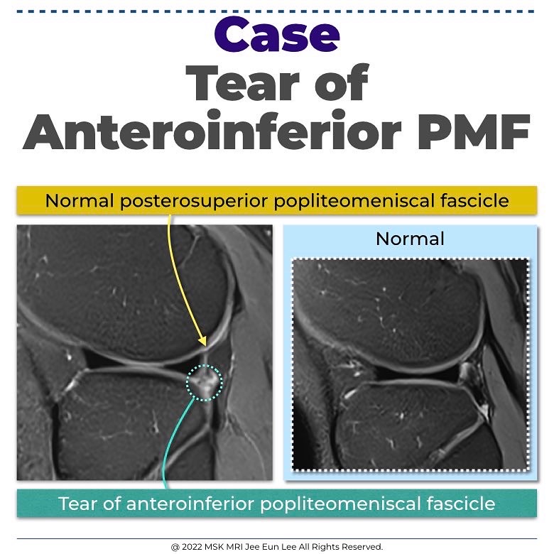
The superior (posterosuperior) and inferior (anteroinferior) popliteomeniscal fascicles can be identified in the coronal and sagittal planes.
A third or posteroinferior popliteomeniscal fascicle is a medial aponeurotic extension from the popliteus musculotendinous area to the inferomedial aspect of the posterior horn of the lateral meniscus.
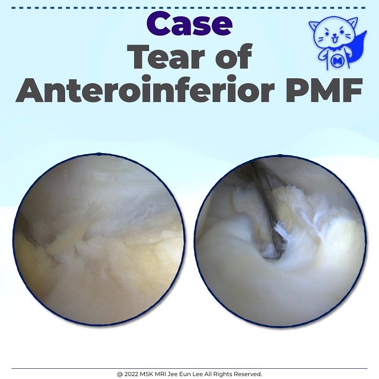
Tearing of the posterosuperior fascicle is highly associated with tears of the posterior horn of the lateral meniscus, with a sensitivity, specificity, and positive predictive value of 89%, 96%, and 79% respectively.
#MSKMRI, #virtualMRI, #radiologist, #musculoskeletal
#KneetMRI,#MSKMRI_Knee
#popliteomeniscal, #lateralmeniscus, #PLCI, #posterolatereralcornerinjury, #PMF
#영상의학과공부맛집, #영상의학, #영상의학이지은, #안산에이스병원,
© 2022 MSK MRI Jee Eun Lee All Rights Reserved.
You may not distribute or commercially exploit the content.
Nor may you transmit it or store it on any other website or other forms of the electronic retrieval system.
Please contact me. If you would like to use images or videos for anything other than personal use.