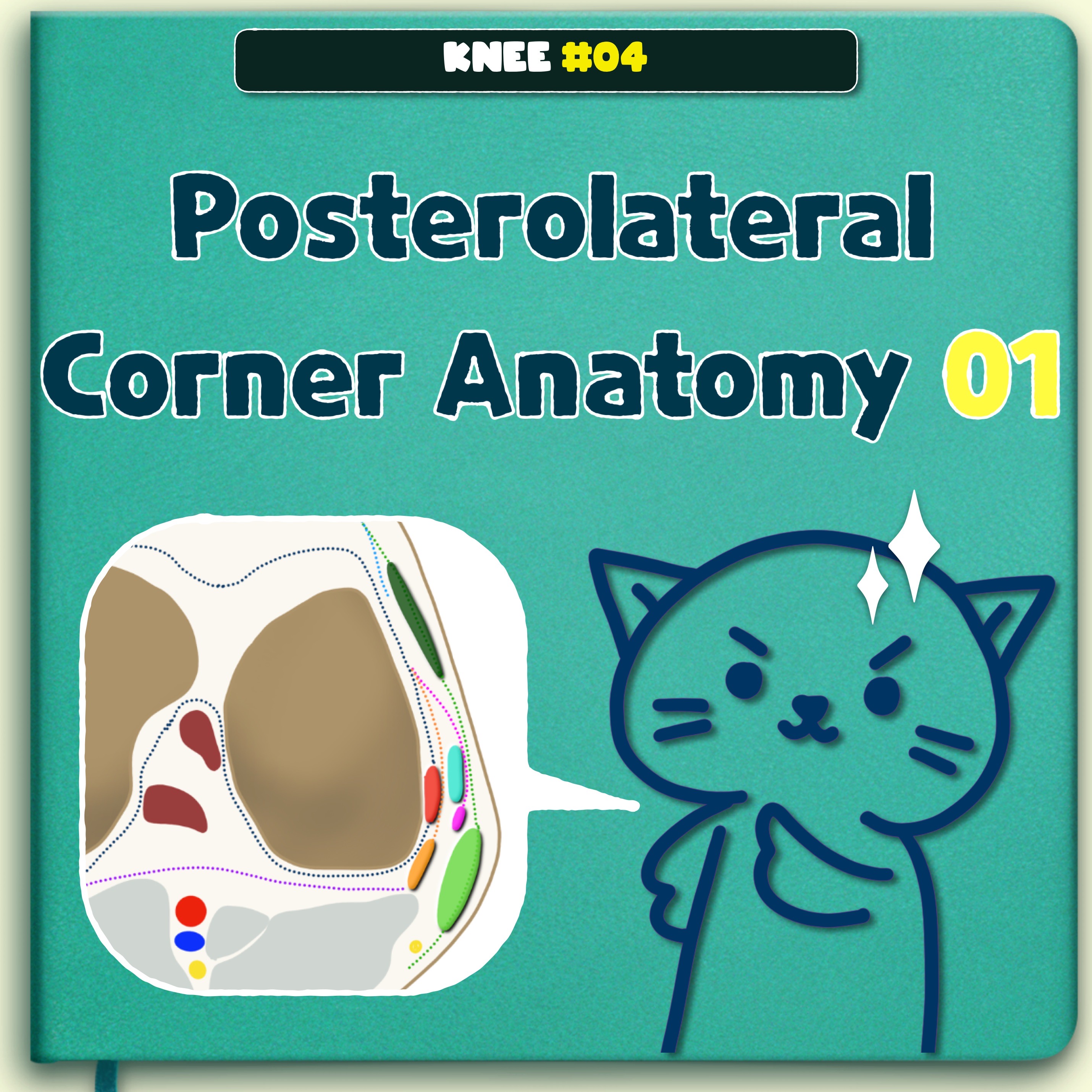
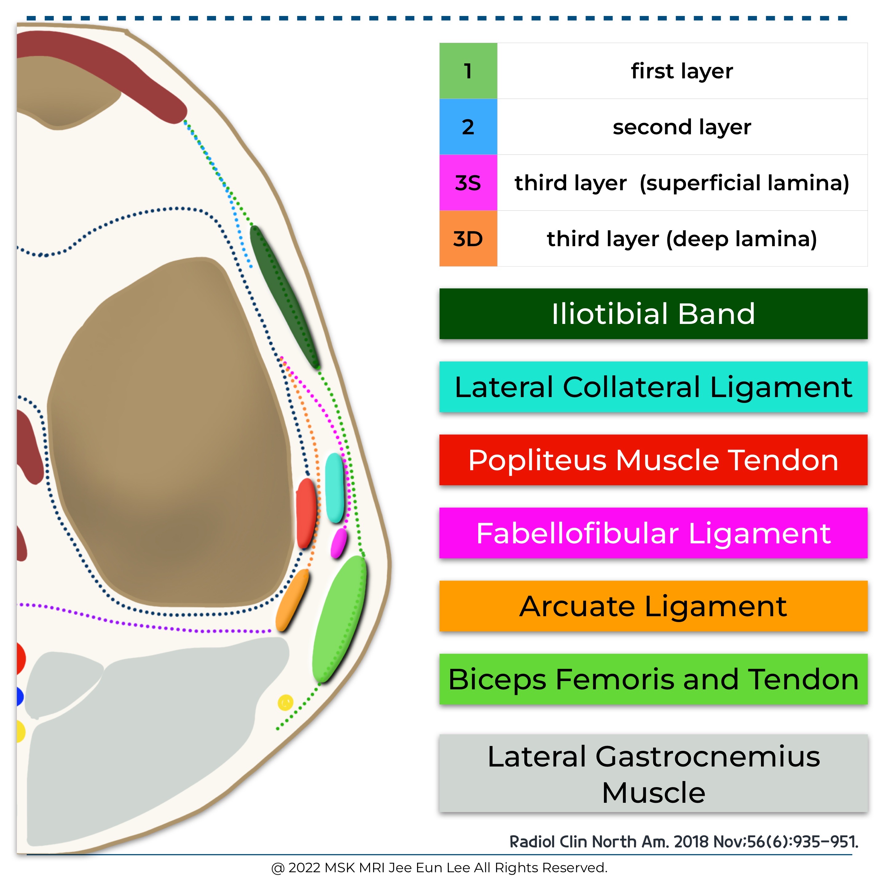
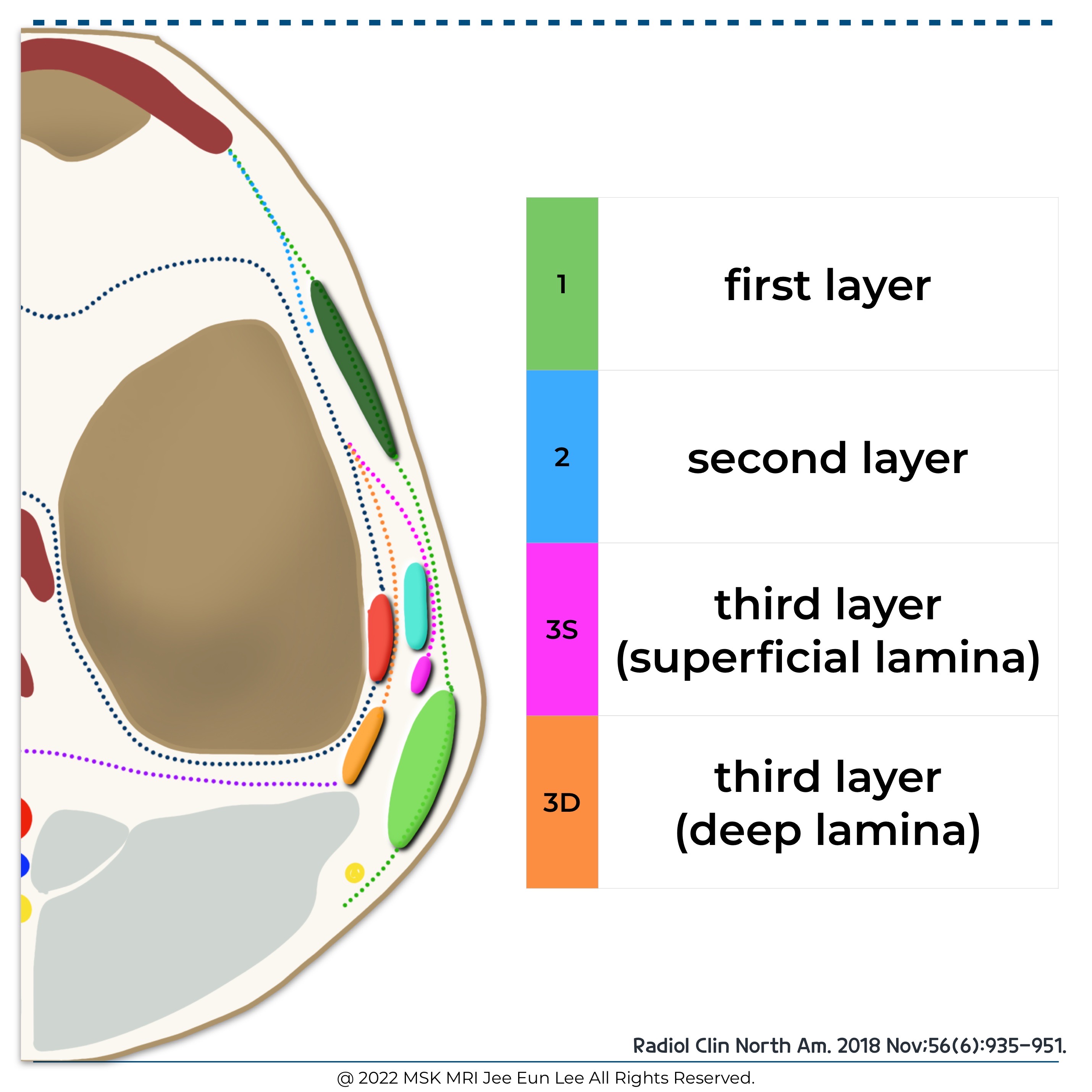
Three structural layers
Layer 1:most superficial
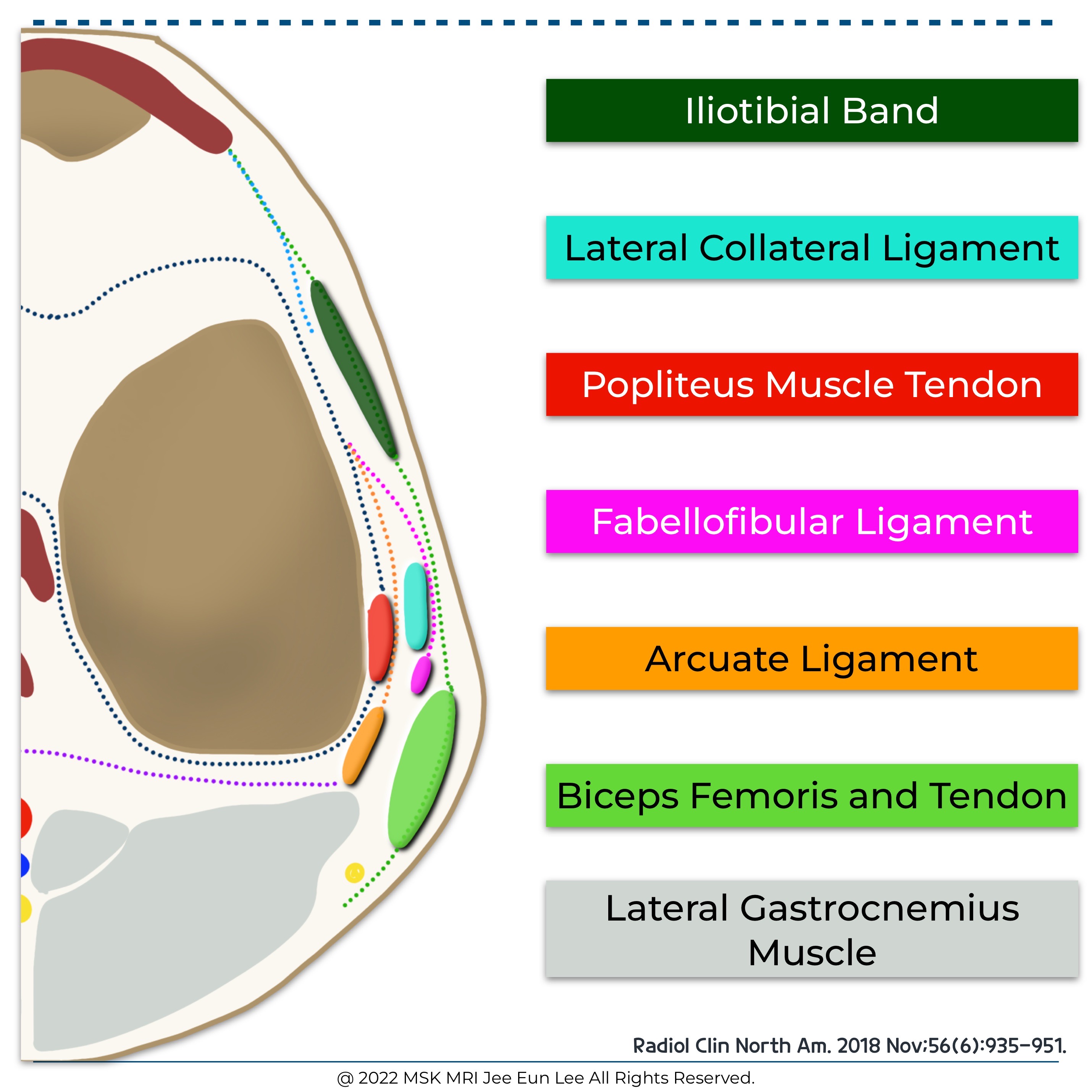
Fascia lata
lliotibial tract with its anterior expansion
Superficial portion of biceps femoris with its posterior expansion
Layer 2
Quadriceps retinaculum anteriorly
Two patellofemoral ligaments or retinacula posteriorly
Lavers 1 and 2 merges at lateral aspect of the patella
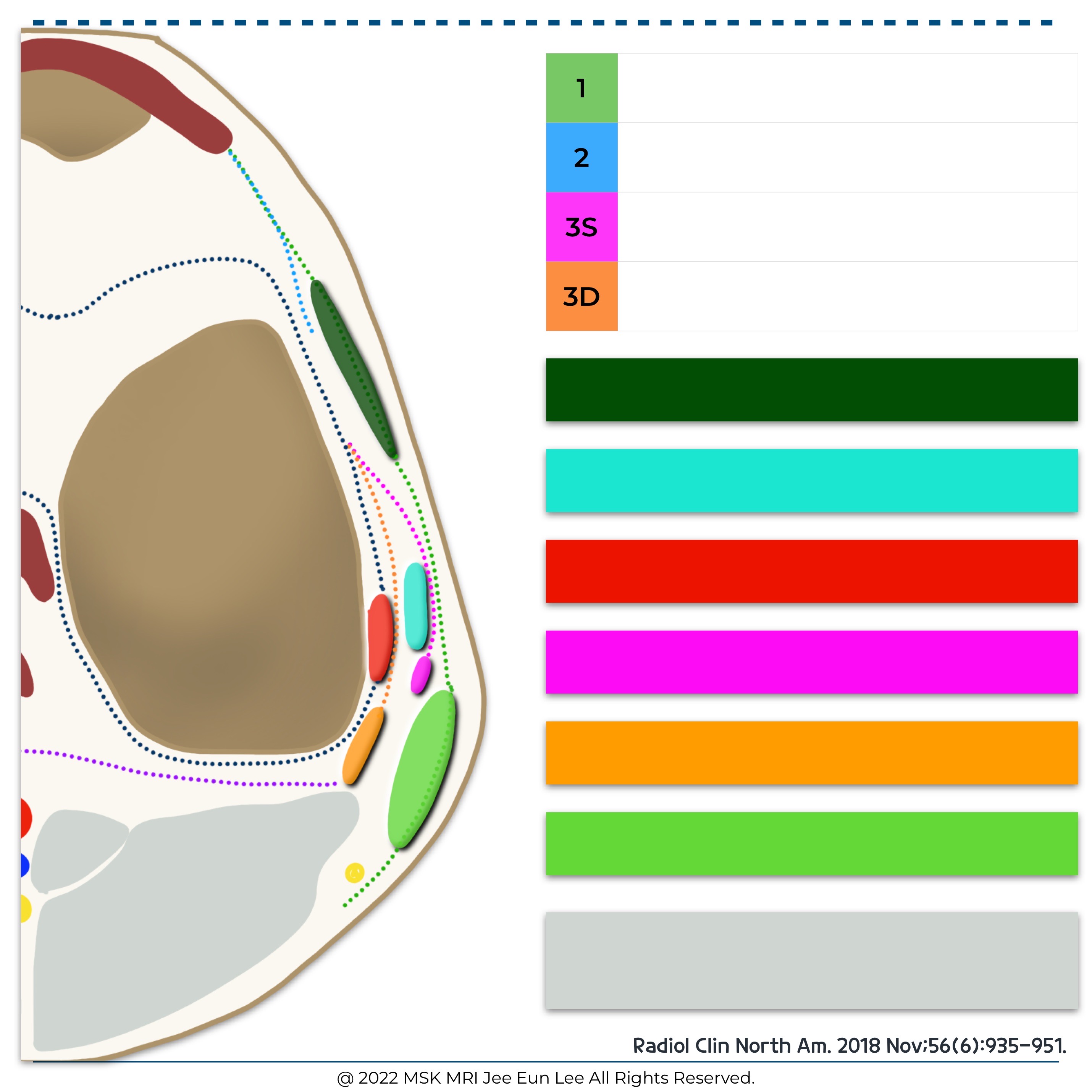
Layer 3:deepest layer
The lateral joint capsule, including attachments to the lateral meniscus
Lateral capsular ligament with meniscofemoral and meniscotibial components
Lateral collateral ligament is located posteriorly between superficial and deep divisions of the layer
Fabellofibular ligament,arcuate ligament,popliteus,and popliteofibulaligament
Deep layer
The deep layer is the most anatomically variable of the 3 layers
The deep and superficial laminae of the posterolateral capsule are always separated from each other with the lateral inferior genicular artery, considered an anatomic landmark, between them
- Superficial lamina of deep layer
- The superficial lamina travels superficial to the lateral collateral ligament and ends posteriorly at the fabellofibular ligament.
- Deeper lamina of deep layer
- The deeper lamina travels deep to the lateral collateral ligament, passes along and attaches to the edge of the lateral meniscus, giving rise to the coronary ligament and ultimately reaching the arcuate ligament.
https://youtube.com/shorts/Az4s4q6NQLA
- YouTube
www.youtube.com
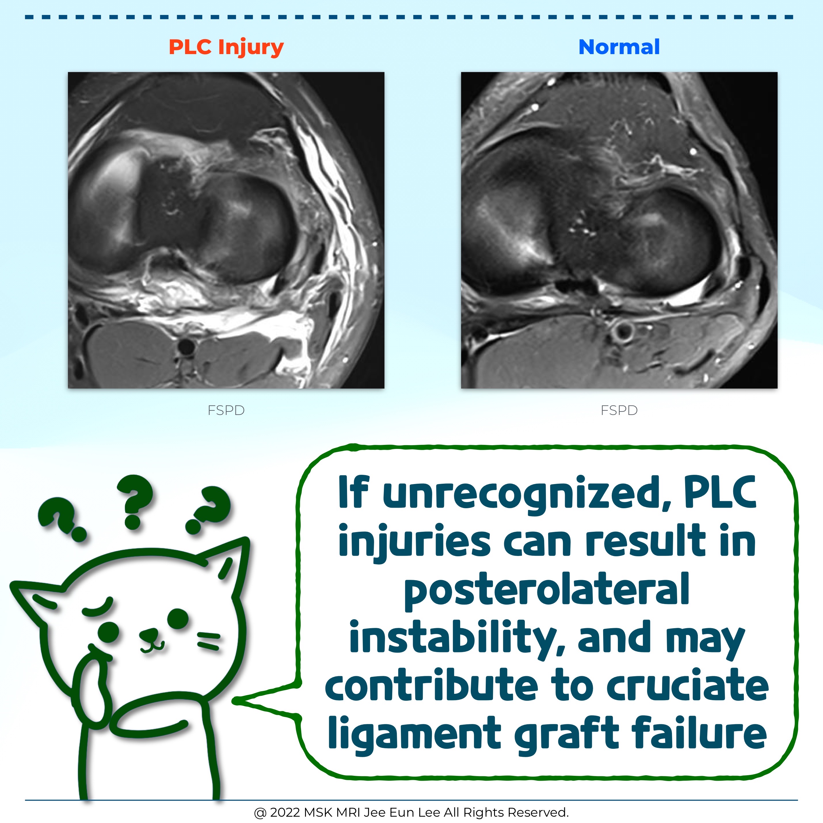
Injury to the arcuate, fabellofibular, or popliteofibular ligament
If edema localizes to the soft tissues of the knee within the PLC, in particular along the superficial aspect of the popliteus, with an otherwise intact biceps femoris, fibular collateral ligament, and popliteus, an injury to the arcuate, fabellofibular, or popliteofibular ligament may be present, especially if these structures cannot be reliably visualized and cleared
The investigators concluded that although MR imaging assessment of the PLC injury is challenging due to anatomic complexity, the larger structures are easy to evaluate with standard MR technique, and direct visualization of small structure involvement on MRI is not necessary to report a clinically unstable PLC injury.
Pearls, Pitfalls, Variants
A recognition of the variations in anatomy and of the limitations of MR imaging with regard to the evaluation of the posterolateral corner (PLC) of the knee is important in the imaging assessment.
The arcuate ligament, fabellofibular ligament, popliteofibular ligament, and popliteomeniscal fascicles are not always perceptible on MR imaging.
An abnormal signal within the region of the posterolateral capsule may imply injury to a smaller structure of the PLC (arcuate ligament, fabellofibular ligament, and popliteofibular ligament).
Radiol Clin North Am. 2018 Nov;56(6):935-951.
What the Referring Physician Needs To Know
Is there injury to the biceps femoris, fibular collateral ligament, or popliteus? If so, is this injury a low-grade partial tear, high grade partial tear, or full-thickness tear?
Is there an injury, or suggestion of injury (such as edema localizing to the posterolateral capsule), to a small structure of the PLC (arcuate ligament, fabellofibular ligament, and popliteofibular ligament)?
Are there additional injuries present involving a cruciate ligament, collateral ligament, hyaline cartilage, or bone?
Radiol Clin North Am. 2018 Nov;56(6):935-951.
#MSKMRI, #JeeEunLeeMSKMRI, #JeeEunLee, #JELMSKMRI,
#virtualMRI, #musculoskeletal,
#영상의학과공부맛집, #영상의학, #영상의학이지은, #안산에이스병원, #ansanacehospital,
© 2022 MSK MRI Jee Eun Lee All Rights Reserved.
모든 컨텐츠의 저작권은 MSK MRI Jee Eun Lee에 있으며, 무단 도용, 배포 및 사용을 금합니다.
사전 협의 없이 무단으로 사용할 경우, 저작권법 등 관련법 위반으로 손해배상 청구 등 민,형사상의 책임과 처벌을 받을 수 있습니다.
You may not distribute or commercially exploit the content.
Nor may you transmit it or store it on any other website or other forms of the electronic retrieval system.
Please contact me. If you would like to use images or videos for anything other than personal use.
(jamaisvu1977@gmail.com)
posterolateral corner injury, popliteus tendon,슬와근, knee mri, anatomy,lateral collateral ligament, fibular collateral ligament, biceps femoris tendon, conjoined tendon, arcuate ligament,fabellofibular ligament,popliteofibular ligament, biceps femoris, posterolateral corner, Iliotibial Band