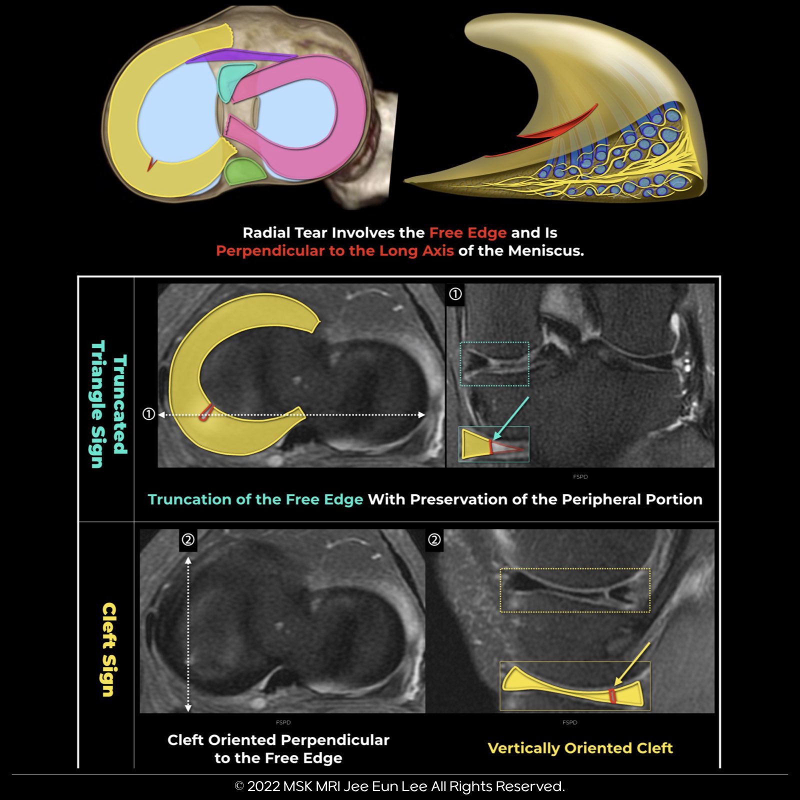
📌 What is a Radial Tear?
• A radial tear runs perpendicular to the tibial plateau and the meniscus’s long axis, transecting the longitudinal collagen bundles.
📌 Significance of Radial Tears:
• Unlike horizontal and longitudinal tears, radial tears significantly disrupt meniscal hoop strength.
• The deeper the tear, the more it impacts the biomechanical function of the menisci.
📍 Common Locations:
• Medial meniscus’s posterior horn.
• Junction of the body and anterior horn in the lateral meniscus.
🔬 Radiologic Signs to Detect Radial Tears:
1. Truncated Triangle Sign:
• Truncation of the free edge, usually indicating a partial-thickness tear.
2. Ghost Meniscus Sign:
• High signal in place of the normally low signal posterior horn, suggesting a full-thickness tear.
3. Cleft Sign:
• Can indicate both longitudinal and radial tears, depending on the tear’s location relative to the imaging plane.
4. Marching Cleft Sign:
• Appears as a progressing cleft away from the free edge on contiguous MR imaging sections, especially at the junction of the horn and body.
💡 Radial tears are crucial in musculoskeletal radiology, affecting the knee’s biomechanical integrity.


#VisualizingMSK #MeniscusTears #MusculoskeletalRadiology
'✅ Knee MRI Mastery > Chap 1. Meniscus' 카테고리의 다른 글
| (Fig 1-B.08) Marching cleft sign in a radial tear (0) | 2024.01.22 |
|---|---|
| (Fig 1-B.07) Full-thickness Radial Meniscal Tears (0) | 2024.01.21 |
| (Fig 1-B.04) Longitudinal-Vertical Meniscal Tears with ACL tear (1) | 2024.01.19 |
| (Fig 1-B.03) Longitudinal-Vertical Meniscal Tears (0) | 2024.01.18 |
| (Fig 1-B.02) Horizontal Meniscal Tears (0) | 2024.01.17 |