Click the link to purchase on Amazon 🎉📚
==============================================
🎥 Check Out All Videos at Once! 📺
👉 Visit Visualizing MSK Blog to explore a wide range of videos! 🩻
https://visualizingmsk.blogspot.com/?view=magazine
📚 You can also find them on MSK MRI Blog and Naver Blog! 📖
https://www.instagram.com/msk_mri/
Click now to stay updated with the latest content! 🔍✨
==============================================
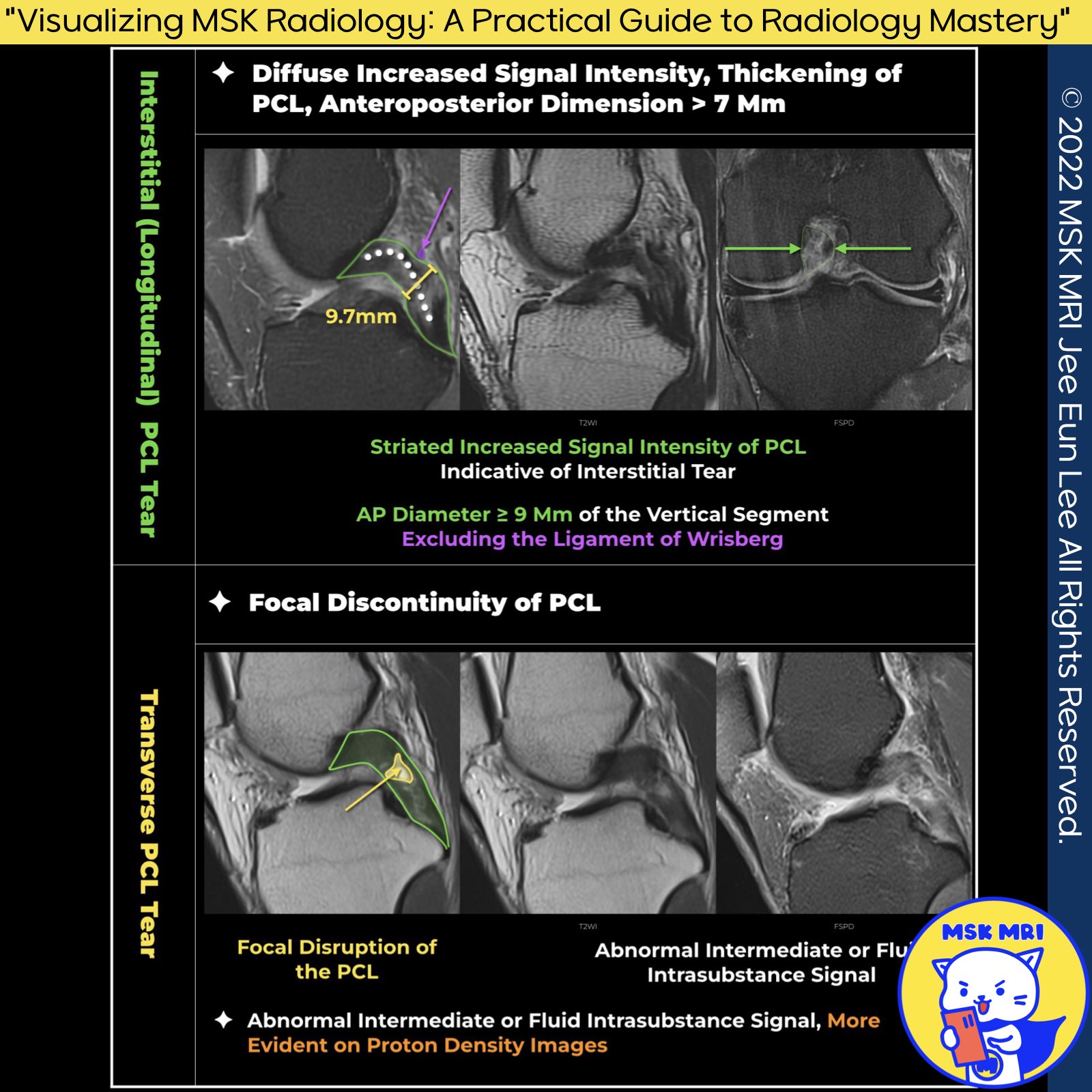
1️⃣ MRI Signs of PCL Tear:
- Focal disruption of the PCL.
- Amorphous increased signal intensity with disruption of one or both margins of the ligament.
- Increased signal intensity on proton density images is more common than on T2-weighted images.
- Nonvisualization of the PCL.
- Abnormal thickening of the PCL distal to the genu with an anteroposterior diameter of greater than 7 mm. (Magn Reson Imaging Clin N Am. 2014 Nov;22(4):557-80.)
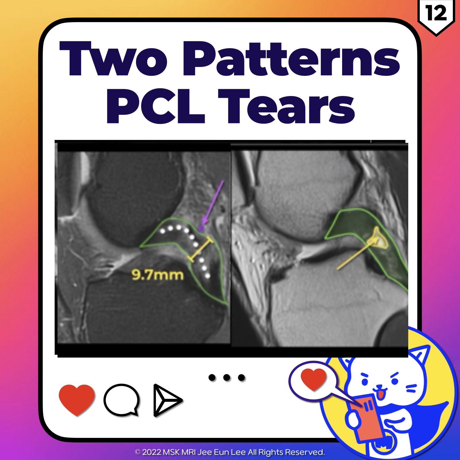
2️⃣ PCL Injury Patterns:
- Injury to the PCL may result in transverse or longitudinal interstitial tears. (Radiology. 2016 Oct;281(1):23-40)
✅ Interstitial Tears: The anteroposterior diameter of the ligament distal to the genu is greater than 7 mm on a sagittal T2-weighted sequence.
✅ Transverse Tears: Focal disruption of the PCL.
https://visualizingmsk.blogspot.com/?view=flipcard
"Visualizing MSK Radiology: A Practical Guide to Radiology Mastery"
© 2022 MSK MRI Jee Eun Lee All Rights Reserved.
#VisualizingMSK #PCLinjuries #KneeMRI #PCLtear
You should not distribute or commercially exploit the content.
You should not transmit or store it on any other website or electronic retrieval system.


'✅ Knee MRI Mastery > Chap 2.ACL and PCL' 카테고리의 다른 글
| (Fig 2-E.14) Common MRI Findings of PCL Tears (0) | 2024.03.16 |
|---|---|
| (Fig 2-E.13) Transverse Tear of PCL And Impingement (0) | 2024.03.16 |
| (Fig 2-E.11) Femoral Periosteal Avulsion Tear Of The PCL (0) | 2024.03.16 |
| (Fig 2-E.10) PCL Ganglia and Mucoid degeneration (0) | 2024.03.16 |
| (Fig 2-E.09) Mucoid Degeneration Of The PCL (0) | 2024.03.16 |