Click the link to purchase on Amazon 🎉📚
==============================================
🎥 Check Out All Videos at Once! 📺
👉 Visit Visualizing MSK Blog to explore a wide range of videos! 🩻
https://visualizingmsk.blogspot.com/?view=magazine
📚 You can also find them on MSK MRI Blog and Naver Blog! 📖
https://www.instagram.com/msk_mri/
Click now to stay updated with the latest content! 🔍✨
==============================================
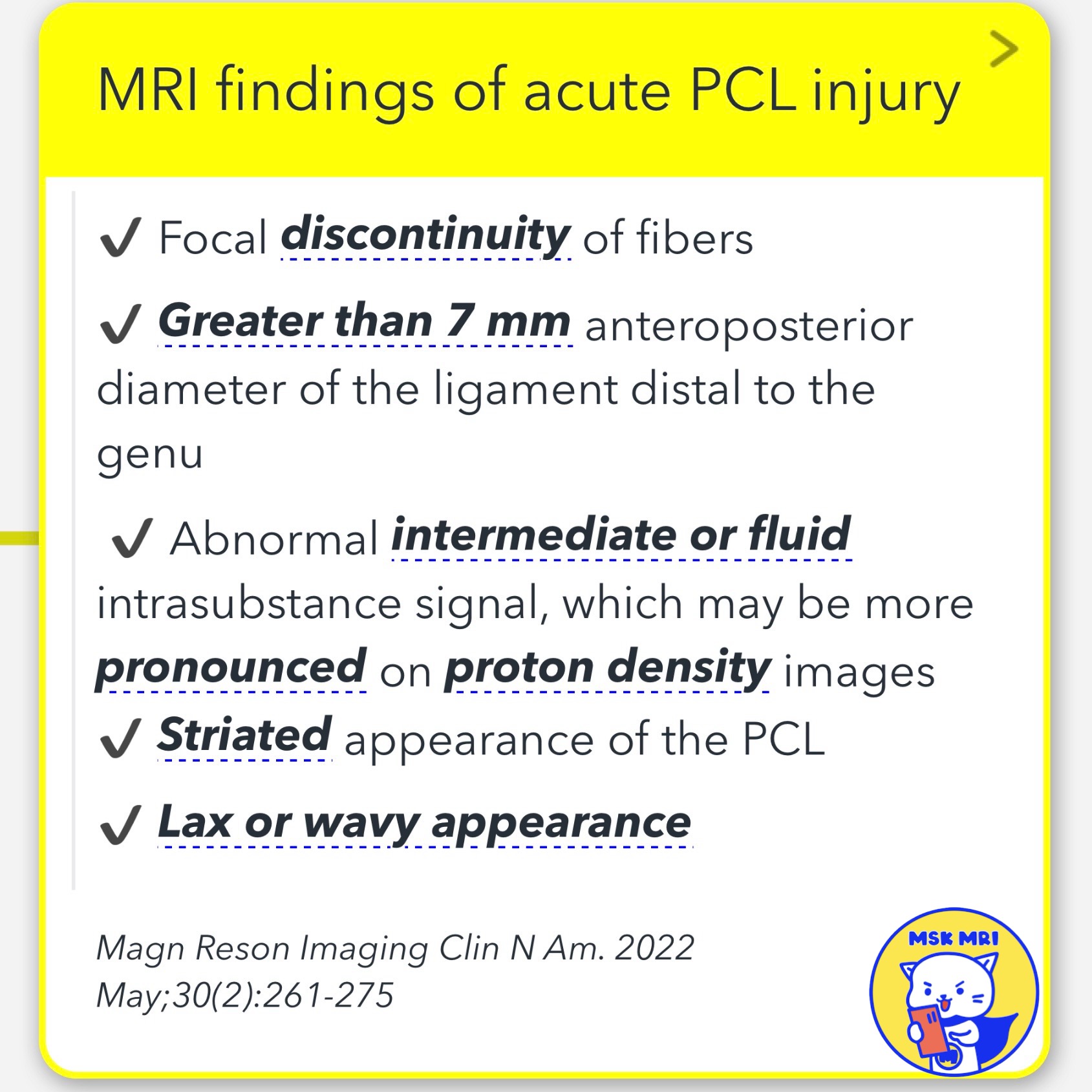
1️⃣ MRI Findings of Acute PCL Injury
✅ Key Indicators:
- Focal discontinuity of fibers.
- The anteroposterior diameter of the ligament distal to the genu is greater than 7 mm.
- Presence of abnormal intermediate or fluid intrasubstance signal, potentially more evident on proton density images.
- Striated appearance of the PCL.
- Lax or wavy appearance of the ligament.
Source: Magnetic Resonance Imaging Clinics of North America. May 2022;30(2):261-275.
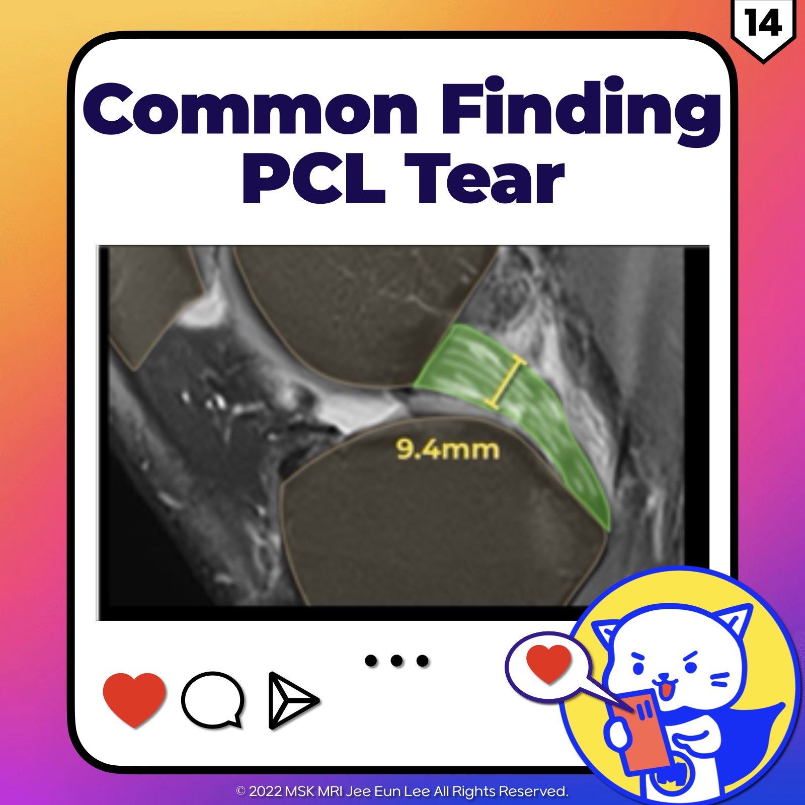
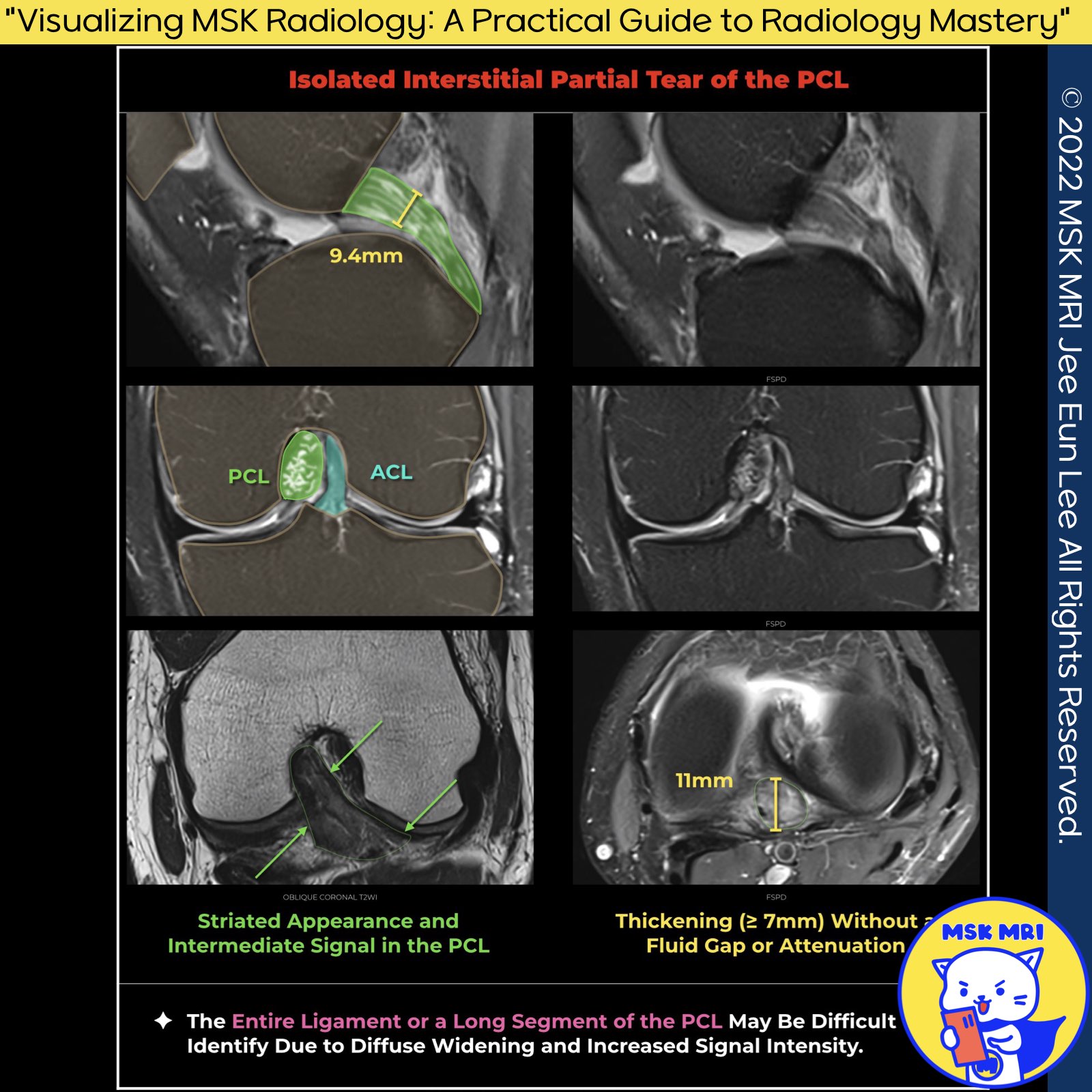
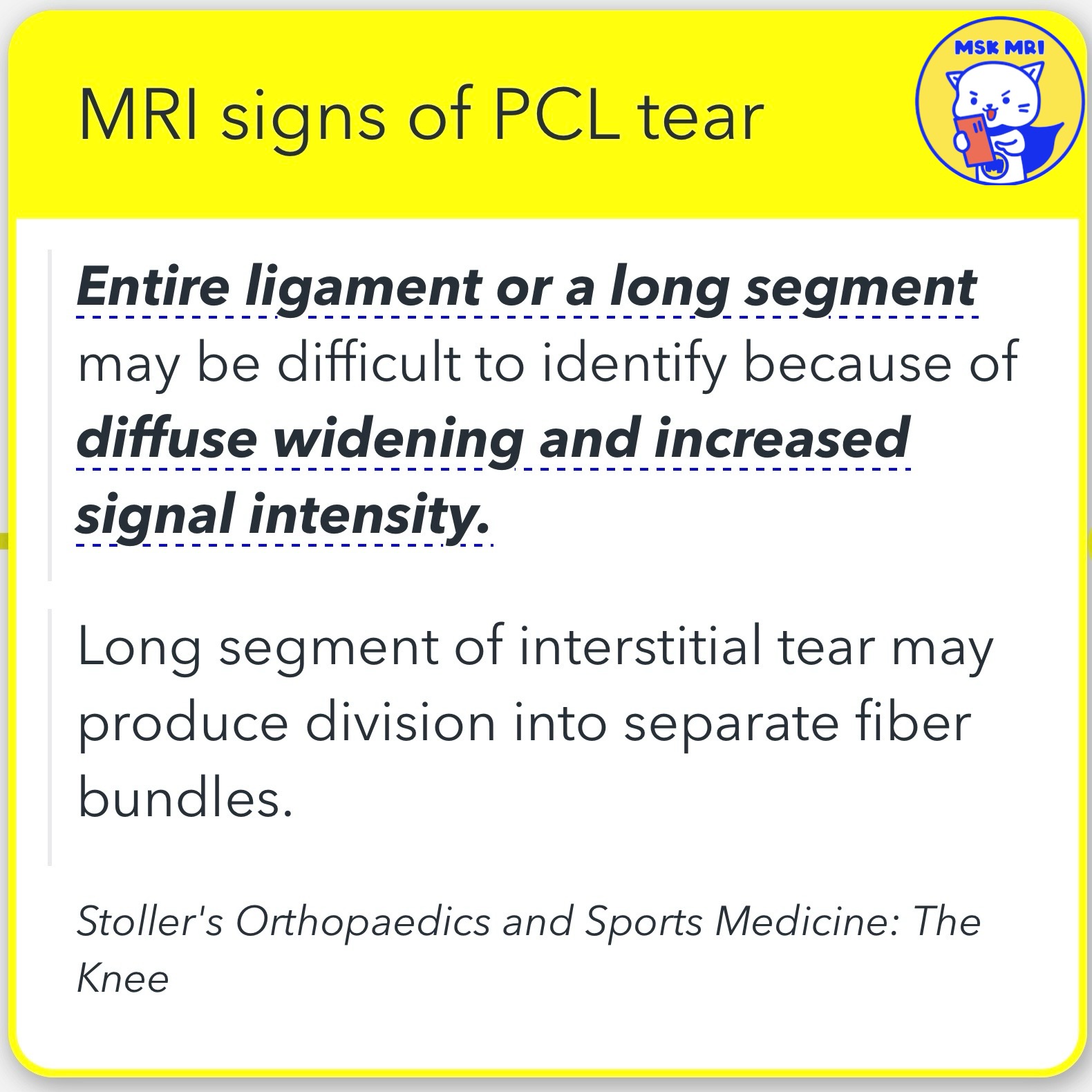
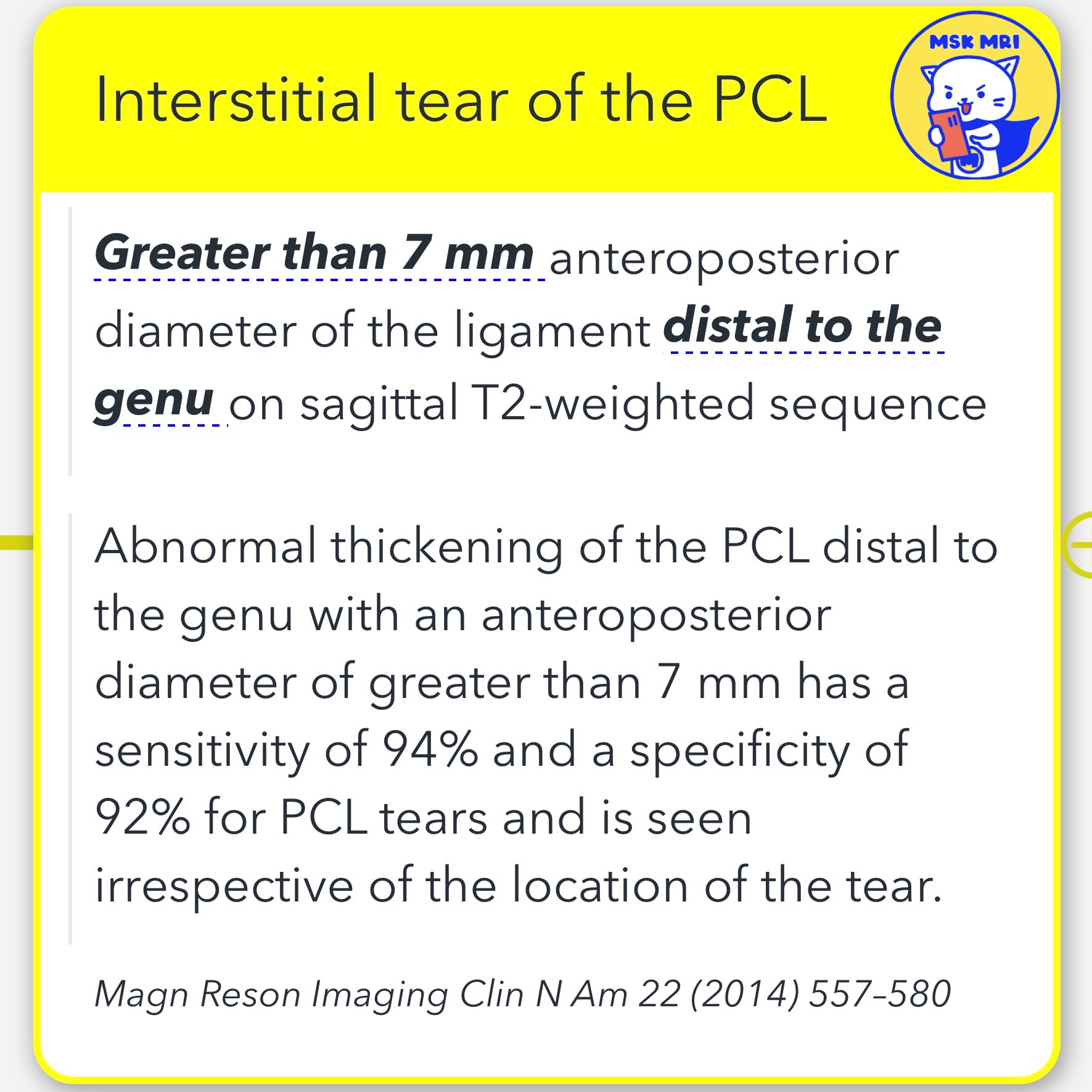
2️⃣ Interstitial Tear of the PCL
✅Diagnostic Criteria:
- A greater than 7 mm anteroposterior diameter of the ligament distal to the genu on sagittal T2-weighted sequence is a key indicator.
- Abnormal thickening of the PCL distal to the genu with an anteroposterior diameter greater than 7 mm has a 94% sensitivity and 92% specificity for PCL tears. This finding is consistent regardless of the tear's location.
- The entire ligament or a long segment may be challenging to identify due to diffuse widening and increased signal intensity.
- A long segment of interstitial tear may result in the division into separate fiber bundles.
Source: Stoller's Orthopaedics and Sports Medicine: The Knee.
Magnetic Resonance Imaging Clinics of North America 22 (2014) 557–580.
https://visualizingmsk.blogspot.com/?view=flipcard
"Visualizing MSK Radiology: A Practical Guide to Radiology Mastery"
© 2022 MSK MRI Jee Eun Lee All Rights Reserved.
#VisualizingMSK #PCLinjuries #KneeMRI #PCLtear
You should not distribute or commercially exploit the content.
You should not transmit or store it on any other website or electronic retrieval system.


'✅ Knee MRI Mastery > Chap 2.ACL and PCL' 카테고리의 다른 글
| (Fig 2-E.17) Partial Tears of PCL and Intact MFL (0) | 2024.03.16 |
|---|---|
| (Fig 2-E.16) Bone Contusions In PCL Hyperextension Injury (2) | 2024.03.16 |
| (Fig 2-E.13) Transverse Tear of PCL And Impingement (0) | 2024.03.16 |
| (Fig 2-E.12) Two Patterns Of The PCL Tears (0) | 2024.03.16 |
| (Fig 2-E.11) Femoral Periosteal Avulsion Tear Of The PCL (0) | 2024.03.16 |